Fetal bone,light micrograph
Numéro d’image : 11711992
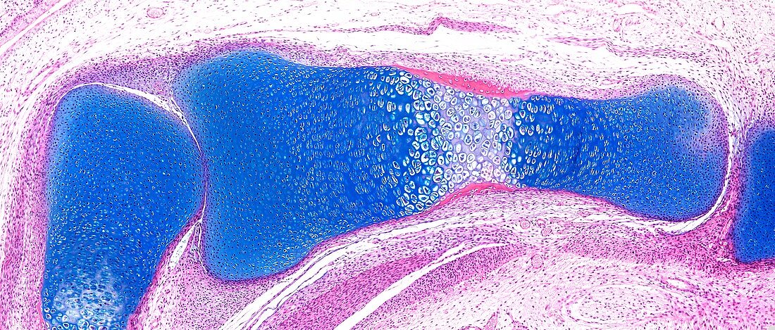
| Light microscopy of a fetal finger bone. At this time of development the future bone is mostly cartilage (blue tissue) with a primary ossification centre in the middle. Here the cartilage cells have enlarged prior to their programmed death that will leave behind a cartilage matrix upon which new bone matrix will be deposited by osteoblasts. A collar of early bone stained red is seen at this central location. Over time most of the cartilage will be replaced with bone leaving a cap of articular cartilage covering the bone ends. This type of bone formation is called endochondral ossification and occurs in almost all of the bones of the axial skeleton and limbs. Magnification x85 when narrow width printed at 10 cm | |
| Licence : | Droits gérés |
| Crédit: | Science Photo Library / Microscape |
| Taille de l’image : | 7087 px × 3019 px |
| Model Release : | Non requis |
| Property Release : | Non requis |
| Restrictions : | - |
Prix pour cette image À partir de 45 €
Produit vendu
(Calendrier, Carte postale, Carte de vœux, Impression sur textile, Packaging etc)
À partir de 45 €
Usage commercial
(Affichage, Annonce presse, Annonce TV, Carte, Digital - hors rés. sociaux, Digital - rés. sociaux etc)
À partir de 45 €
Éditorial
(Digital, Journal, Livre, Livre pratique, Magazine, Télévision etc)
À partir de 60 €
Usage non-commercial
(Digital - hors rés. sociaux, Digital - rés. sociaux etc)
À partir de 120 €
Mots clés
- biologie,
- biologique,
- cartilage,
- catégorie,
- centre ossification,
- chondrocytes,
- coupe,
- developpement des os,
- développement du squelette,
- développement squelette,
- développement squelettique,
- développment osseux,
- histologie,
- histologique,
- matrice du cartilage,
- microscope optique,
- microscope photonique,
- microscopie optique,
- microscopie photonique,
- os,
- ossification,
- partie,
- sclérose,
- section,
- squelette,
- squelettique