Spinal nerve ganglion,light micrograph
Numéro d’image : 11711984
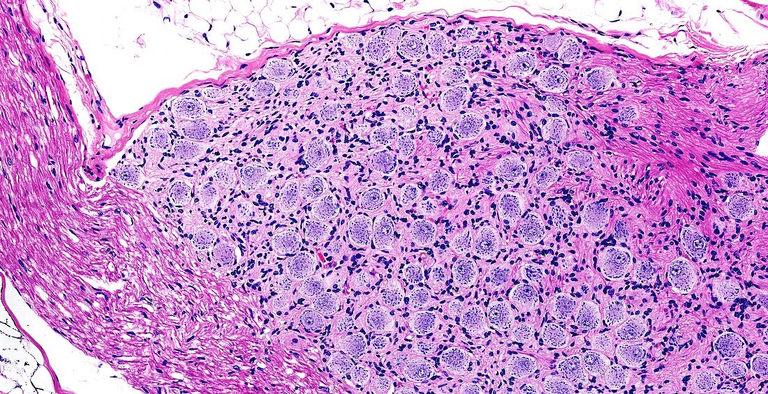
| Light microscopy of a sensory spinal nerve ganglion also called a dorsal root ganglion. It consists of nerve cell bodies (blue) accompanied by many small satellite or glial cells. The pink-stained tissues are myelinated nerves entering and leaving the ganglion. Sensory signals from peripheral organs including skin travel via nerves towards the spinal cord and pass through the ganglion before entering the grey matter (dorsal horn) of the spinal cord. Magnification x170 when printed at 10cm | |
| Licence : | Droits gérés |
| Crédit: | Science Photo Library / Microscape |
| Taille de l’image : | 5906 px × 3033 px |
| Model Release : | Non requis |
| Property Release : | Non requis |
| Restrictions : | - |
Prix pour cette image À partir de 45 €
Produit vendu
(Calendrier, Carte postale, Carte de vœux, Impression sur textile, Packaging etc)
À partir de 45 €
Usage commercial
(Affichage, Annonce presse, Annonce TV, Carte, Digital - hors rés. sociaux, Digital - rés. sociaux etc)
À partir de 45 €
Éditorial
(Digital, Journal, Livre, Livre pratique, Magazine, Télévision etc)
À partir de 60 €
Usage non-commercial
(Digital - hors rés. sociaux, Digital - rés. sociaux etc)
À partir de 120 €