Skin naevus,light micrograph
Numéro d’image : 11711982
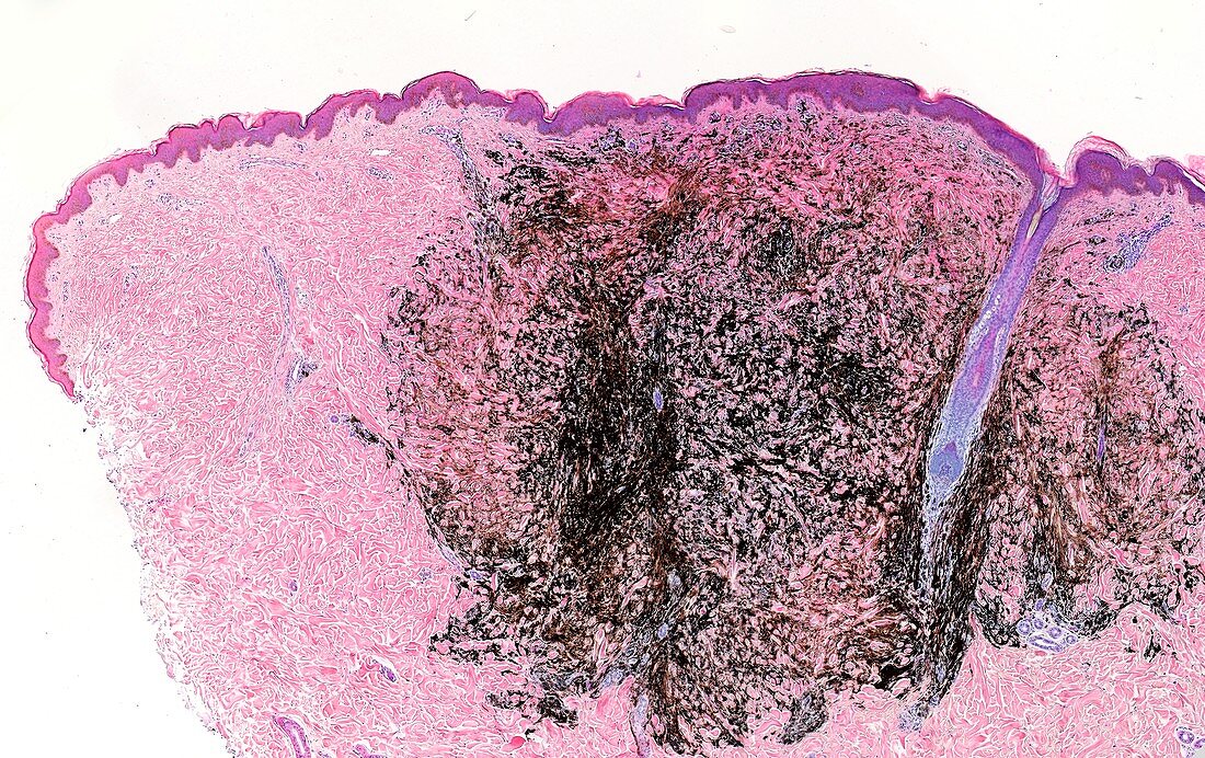
| Light microscopy of a skin naevus sometimes referred to as a mole. The naevus shows a abnormal proliferation of pigment-producing cells melanocytes. This is a deep penetrating naevus with extensive growth into the dermis. The fine cytoplasmic extensions of many thousands of melanocytes are filled with dark pigment granules giving a dense network pattern of pigment. Naevi are usually benign . The macroscopic appearance may give the classification as a blue naevus. Magnification x30 when printed at 10 cm | |
| Licence : | Droits gérés |
| Crédit: | Science Photo Library / Microscape |
| Taille de l’image : | 5315 px × 3346 px |
| Model Release : | Non requis |
| Property Release : | Non requis |
| Restrictions : | - |
Prix pour cette image À partir de 45 €
Produit vendu
(Calendrier, Carte postale, Carte de vœux, Impression sur textile, Packaging etc)
À partir de 45 €
Usage commercial
(Affichage, Annonce presse, Annonce TV, Carte, Digital - hors rés. sociaux, Digital - rés. sociaux etc)
À partir de 45 €
Éditorial
(Digital, Journal, Livre, Livre pratique, Magazine, Télévision etc)
À partir de 60 €
Usage non-commercial
(Digital - hors rés. sociaux, Digital - rés. sociaux etc)
À partir de 120 €