Testis,light micrograph
Numéro d’image : 11704791
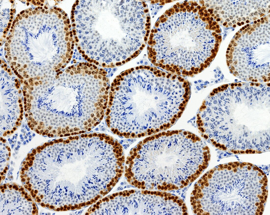
| Light microscopy of the testis. Seminiferous tubules are shown in cross-section. Tubules contain the seminiferous epithelium where germ cells multiply and develop into sperm. Sperm heads and tails stain bright blue. Cells showing DNA synthesis (spermatogonia,early primary spermatocytes) have been selectively stained brown with an immunostaining technique for proliferating cell nuclear antigen (PCNA). This stain demonstrates that DNA replication is restricted to mitotic spermatogonia and early meiotic spermatocytes. All other germ cells are not engaged in DNA synthesis. Magnification x150 when printed at 10 cm | |
| Licence : | Droits gérés |
| Crédit: | Science Photo Library / Microscape |
| Taille de l’image : | 4710 px × 3748 px |
| Model Release : | Non requis |
| Property Release : | Non requis |
| Restrictions : | - |
Prix pour cette image À partir de 45 €
Produit vendu
(Calendrier, Carte postale, Carte de vœux, Impression sur textile, Packaging etc)
À partir de 45 €
Usage commercial
(Affichage, Annonce presse, Annonce TV, Carte, Digital - hors rés. sociaux, Digital - rés. sociaux etc)
À partir de 45 €
Éditorial
(Digital, Journal, Livre, Livre pratique, Magazine, Télévision etc)
À partir de 60 €
Usage non-commercial
(Digital - hors rés. sociaux, Digital - rés. sociaux etc)
À partir de 120 €
Mots clés
- ADN,
- biologie,
- biologique,
- catégorie,
- cellules germinales,
- coupe,
- épithélium séminifère,
- germen,
- histologie,
- histologique,
- méiose,
- microscope optique,
- microscopie optique,
- mitose,
- partie,
- PCNA,
- section,
- spermatocytes,
- spermatogenèse,
- spermatogonie,
- synthèse ADN,
- testicules,
- testis,
- tubules séminifères