Tooth,light micrograph
Numéro d’image : 11704780
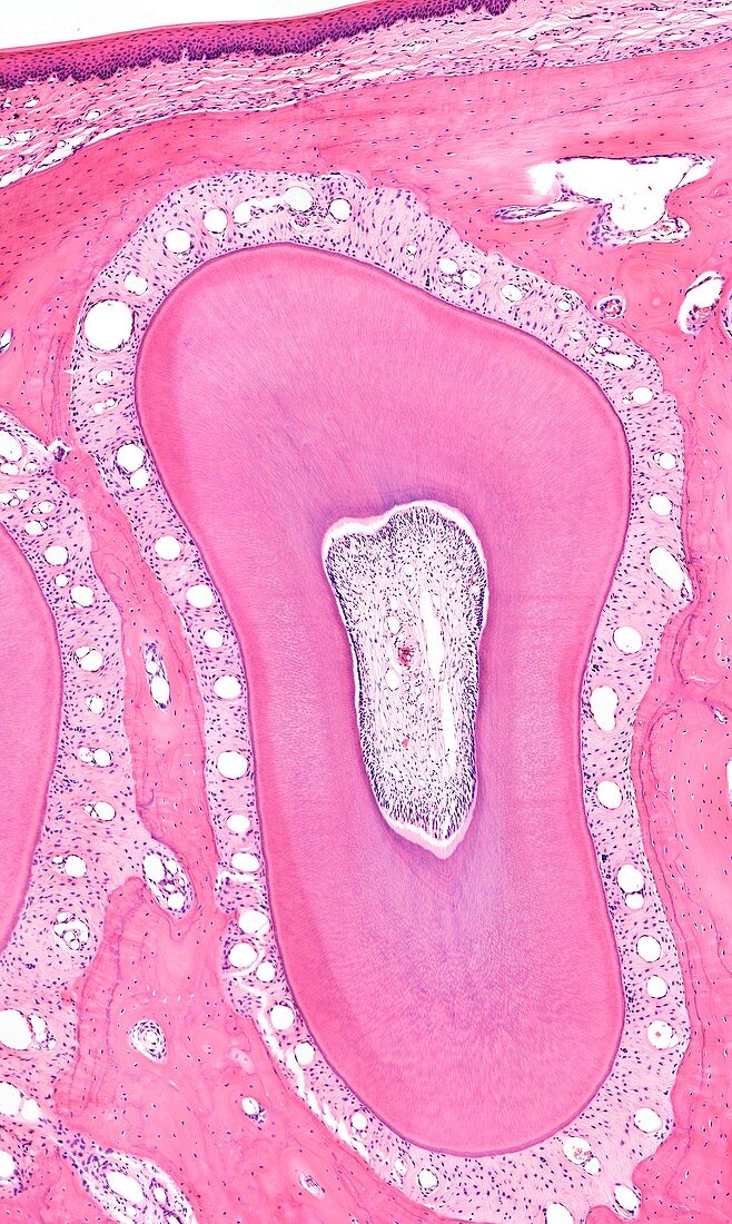
| Light microscopy of a tooth. This section is below the gumline and shows how the root of the tooth is bordered by a connective tissue zone called the periodontal ligament. It suspends the tooth in a socket of jaw bone stained pink. Spaces in the ligament are blood vessels and nerves are also present. The centre of the tooth is the pulp cavity (root canal) that also has vessels and nerves. Most of the tooth seen here is dentin,a hard tissue consisting of fine,long tubules that contain collagen,proteins and mineralized matrix. Magnification x75 when printed at 10 cm | |
| Licence : | Droits gérés |
| Crédit: | Science Photo Library / Microscape |
| Taille de l’image : | 3245 px × 5422 px |
| Model Release : | Non requis |
| Property Release : | Non requis |
| Restrictions : | - |
Prix pour cette image À partir de 45 €
Produit vendu
(Calendrier, Carte postale, Carte de vœux, Impression sur textile, Packaging etc)
À partir de 45 €
Usage commercial
(Affichage, Annonce presse, Annonce TV, Carte, Digital - hors rés. sociaux, Digital - rés. sociaux etc)
À partir de 45 €
Éditorial
(Digital, Journal, Livre, Livre pratique, Magazine, Télévision etc)
À partir de 60 €
Usage non-commercial
(Digital - hors rés. sociaux, Digital - rés. sociaux etc)
À partir de 120 €