Peripheral nerve,light micrograph
Numéro d’image : 11704772
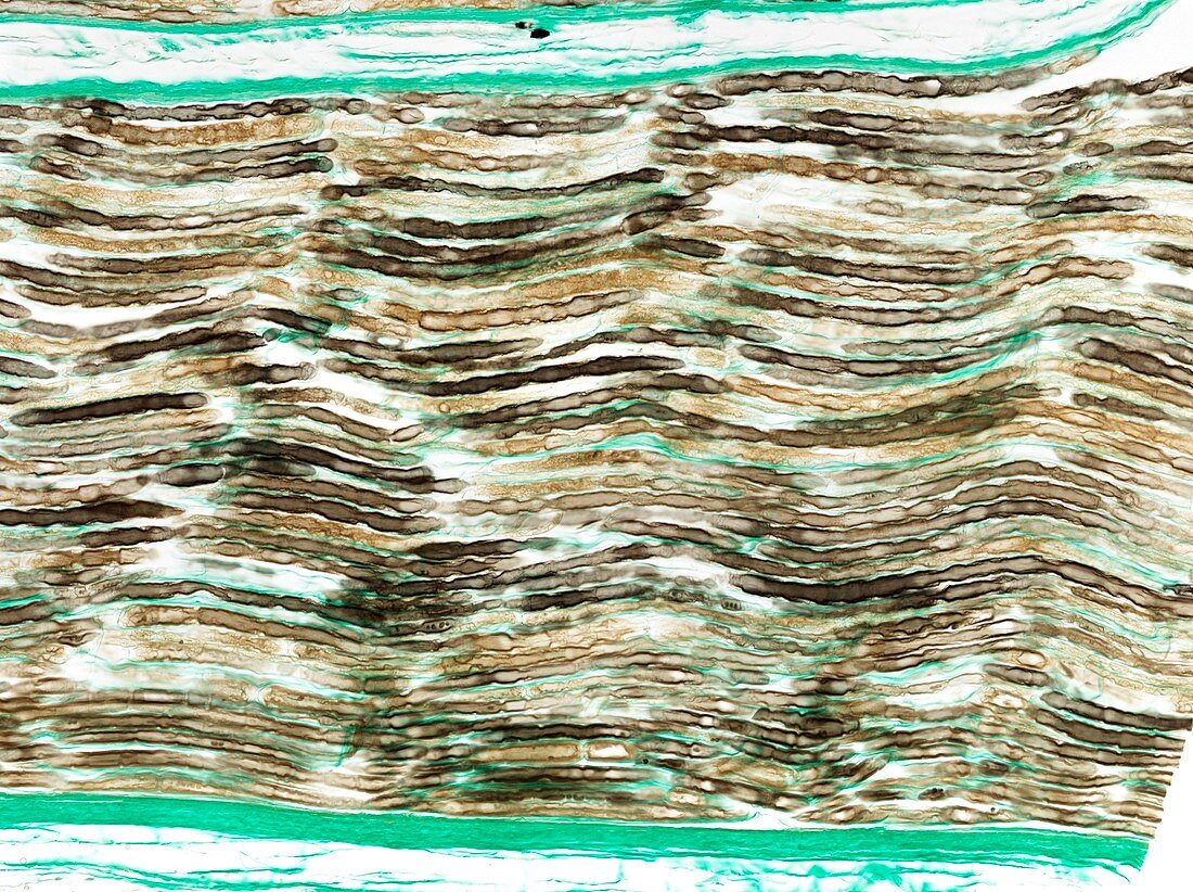
| Light microscopy of myelinated nerves in a nerve fibre. Axons appear as tubular profiles with sharp borders representing myelin sheaths produced by Schwann cells. At intervals along the individual nerves are many constrictions. These are called nodes of Ranvier and signify a small segment where myelin is absent. The myelin sheath between two nodes is produced by a single Schwann cell. Electrical signal conduction by myelinated nerves jumps very rapidly from node to node. Magnification x80 when printed at 10 cm | |
| Licence : | Droits gérés |
| Crédit: | Science Photo Library / Microscape |
| Taille de l’image : | 4859 px × 3632 px |
| Model Release : | Non requis |
| Property Release : | Non requis |
| Restrictions : | - |
Prix pour cette image À partir de 45 €
Produit vendu
(Calendrier, Carte postale, Carte de vœux, Impression sur textile, Packaging etc)
À partir de 45 €
Usage commercial
(Affichage, Annonce presse, Annonce TV, Carte, Digital - hors rés. sociaux, Digital - rés. sociaux etc)
À partir de 45 €
Éditorial
(Digital, Journal, Livre, Livre pratique, Magazine, Télévision etc)
À partir de 60 €
Usage non-commercial
(Digital - hors rés. sociaux, Digital - rés. sociaux etc)
À partir de 120 €