Small bowel,light micrograph
Numéro d’image : 11704768
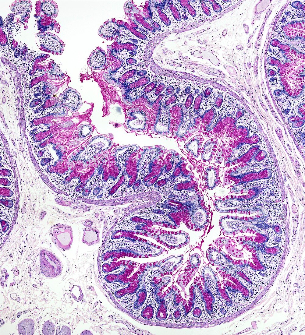
| Light microscopy of the inner lining (mucosa) of the small bowel. The surface consists mainly of fluid-absorbing epithelial cells (blue) and larger mucus-secreting goblet cells (red) that together form finger-like extensions called villi. Blind-ending tubular glands called crypts of Lieberkuhn extend deeply between the villi. The villi increase the inner surface area of the bowel for absorptive and secretory functions. Magnification x90 when narrow width printed at 10 cm | |
| Licence : | Droits gérés |
| Crédit: | Science Photo Library / Microscape |
| Taille de l’image : | 4006 px × 4403 px |
| Model Release : | Non requis |
| Property Release : | Non requis |
| Restrictions : | - |
Prix pour cette image À partir de 45 €
Produit vendu
(Calendrier, Carte postale, Carte de vœux, Impression sur textile, Packaging etc)
À partir de 45 €
Usage commercial
(Affichage, Annonce presse, Annonce TV, Carte, Digital - hors rés. sociaux, Digital - rés. sociaux etc)
À partir de 45 €
Éditorial
(Digital, Journal, Livre, Livre pratique, Magazine, Télévision etc)
À partir de 60 €
Usage non-commercial
(Digital - hors rés. sociaux, Digital - rés. sociaux etc)
À partir de 120 €
Mots clés
- biologie,
- biologique,
- catégorie,
- cellule à mucus,
- cellule caliciforme,
- cellule en gobelet,
- cellule mucipare,
- cellule muqueuse à pôle apical ouvert,
- coupe,
- crypte Lieberkuhn,
- gut,
- histologie,
- histologique,
- intestin,
- intestin grêle,
- microscope optique,
- microscopie optique,
- partie,
- petit intestin,
- section,
- système gastro-intestinal,
- tractus gastro-intestinal,
- villosités