Testis,light micrograph
Numéro d’image : 11704766
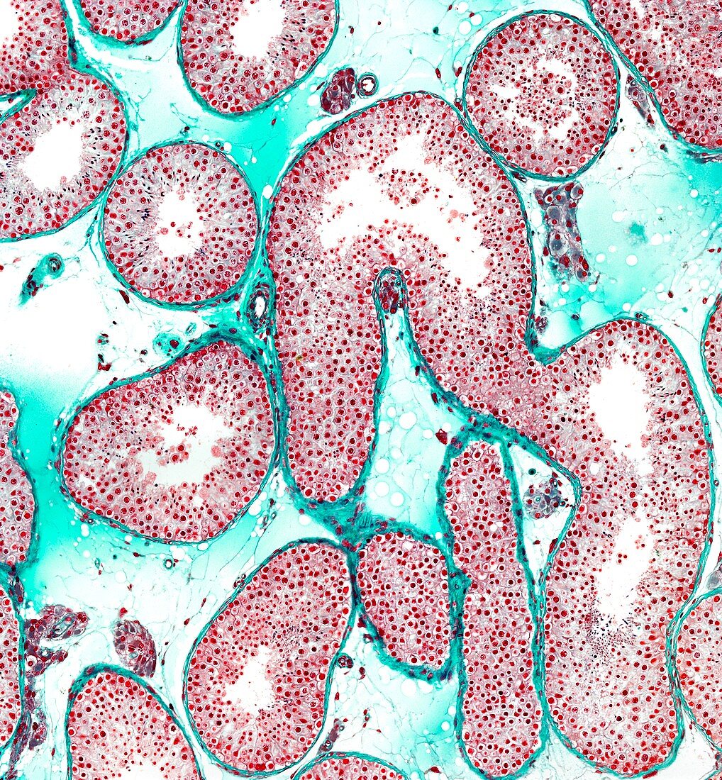
| Light microscopy of the human testis. Seminiferous tubules are shown in section. Tubules are bordered by a well-defined peritubular tissue and they contain male germ cells and non-germinal Sertoli cells making up the seminiferous epithelium. Through the process of spermatogenesis,germ cells multiply and mature over a period of 70 days to produce spermatozoa. Between the tubules clusters of Leydig cells are noted. These cells secrete testosterone which stimulates spermatogenesis. Magnification x60 when printed at 10 cm | |
| Licence : | Droits gérés |
| Crédit: | Science Photo Library / Microscape |
| Taille de l’image : | 4041 px × 4367 px |
| Model Release : | Non requis |
| Property Release : | Non requis |
| Restrictions : | - |
Prix pour cette image À partir de 45 €
Produit vendu
(Calendrier, Carte postale, Carte de vœux, Impression sur textile, Packaging etc)
À partir de 45 €
Usage commercial
(Affichage, Annonce presse, Annonce TV, Carte, Digital - hors rés. sociaux, Digital - rés. sociaux etc)
À partir de 45 €
Éditorial
(Digital, Journal, Livre, Livre pratique, Magazine, Télévision etc)
À partir de 60 €
Usage non-commercial
(Digital - hors rés. sociaux, Digital - rés. sociaux etc)
À partir de 120 €
Mots clés
- biologie,
- biologique,
- catégorie,
- cellule de Leydig,
- coupe,
- épithélium séminifère,
- histologie,
- histologique,
- humain,
- microscope optique,
- microscopie optique,
- organe masculin,
- organe reproducteur,
- organe reproducteur masculin,
- partie,
- section,
- spermatogenèse,
- spermatozoïde,
- testicules,
- testis,
- tube séminifère