Testis,light micrograph
Numéro d’image : 11704765
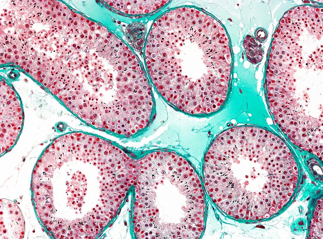
| Light microscopy of the human testis. Seminiferous tubules are shown in section. Tubules are bordered by a well-defined peritubular tissue and they contain male germ cells and non-germinal Sertoli cells making up the seminiferous epithelium. Through the process of spermatogenesis,germ cells multiply and mature over a period of 70 days to produce spermatozoa. Between some of the tubules clusters of Leydig cells are noted. These cells secrete testosterone which stimulates spermatogenesis. Magnification x60 when printed at 10 cm | |
| Licence : | Droits gérés |
| Crédit: | Science Photo Library / Microscape |
| Taille de l’image : | 4864 px × 3598 px |
| Model Release : | Non requis |
| Property Release : | Non requis |
| Restrictions : | - |
Prix pour cette image À partir de 45 €
Produit vendu
(Calendrier, Carte postale, Carte de vœux, Impression sur textile, Packaging etc)
À partir de 45 €
Usage commercial
(Affichage, Annonce presse, Annonce TV, Carte, Digital - hors rés. sociaux, Digital - rés. sociaux etc)
À partir de 45 €
Éditorial
(Digital, Journal, Livre, Livre pratique, Magazine, Télévision etc)
À partir de 60 €
Usage non-commercial
(Digital - hors rés. sociaux, Digital - rés. sociaux etc)
À partir de 120 €
Mots clés
- biologie,
- biologique,
- catégorie,
- cellule de Leydig,
- coupe,
- épithélium séminifère,
- histologie,
- histologique,
- humain,
- microscope optique,
- microscopie optique,
- organe masculin,
- organe reproducteur,
- organe reproducteur masculin,
- partie,
- section,
- spermatogenèse,
- spermatozoïde,
- testicules,
- testis,
- tube séminifère