Peripheral nerve,light micrograph
Numéro d’image : 11704759
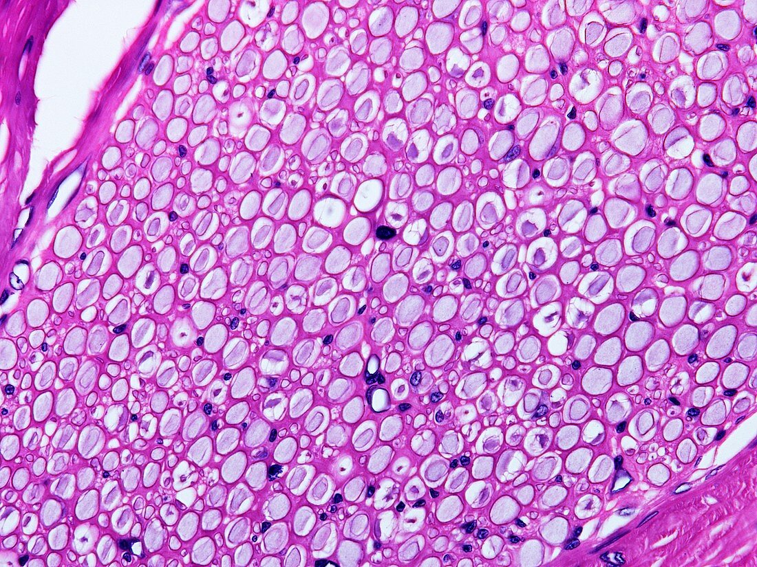
| Light microscopy of myelinated nerves in a cross-sectioned nerve fibre. Axons appear as circular or elliptical profiles enclosed by white rings representing myelin sheaths produced by Schwann cells. Schwann cell nuclei are occasionally seen close to an axon but most of the cell nuclei (dark blue) are fibroblasts of the connective tissues (pink) that support the nerve fibre. Magnification x250 when printed at 10 cm | |
| Licence : | Droits gérés |
| Crédit: | Science Photo Library / Microscape |
| Taille de l’image : | 4828 px × 3621 px |
| Model Release : | Non requis |
| Property Release : | Non requis |
| Restrictions : | - |
Prix pour cette image À partir de 45 €
Produit vendu
(Calendrier, Carte postale, Carte de vœux, Impression sur textile, Packaging etc)
À partir de 45 €
Usage commercial
(Affichage, Annonce presse, Annonce TV, Carte, Digital - hors rés. sociaux, Digital - rés. sociaux etc)
À partir de 45 €
Éditorial
(Digital, Journal, Livre, Livre pratique, Magazine, Télévision etc)
À partir de 60 €
Usage non-commercial
(Digital - hors rés. sociaux, Digital - rés. sociaux etc)
À partir de 120 €