Bone and cartilage,light micrograph
Numéro d’image : 11704753
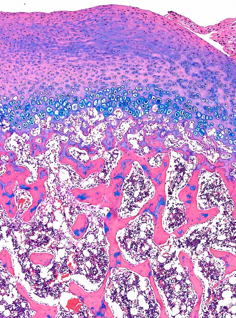
| Light microscopy of developing bone. The top layer (purple,pink) is cartilage which covers the end of the bone; next layer (blue) is the epiphyseal growth plate comprising chondrocytes; lower half shows spongy bone (pink) of honeycomb appearance with remnants of calcifying cartilage (blue) surrounded by bone tissue. The gaps between the spongy bone is occupied by bone marrow cells. Magnification x90 when narrow width printed at 10 cm | |
| Licence : | Droits gérés |
| Crédit: | Science Photo Library / Microscape |
| Taille de l’image : | 3629 px × 4890 px |
| Model Release : | Non requis |
| Property Release : | Non requis |
| Restrictions : | - |
Prix pour cette image À partir de 45 €
Produit vendu
(Calendrier, Carte postale, Carte de vœux, Impression sur textile, Packaging etc)
À partir de 45 €
Usage commercial
(Affichage, Annonce presse, Annonce TV, Carte, Digital - hors rés. sociaux, Digital - rés. sociaux etc)
À partir de 45 €
Éditorial
(Digital, Journal, Livre, Livre pratique, Magazine, Télévision etc)
À partir de 60 €
Usage non-commercial
(Digital - hors rés. sociaux, Digital - rés. sociaux etc)
À partir de 120 €