Spinal sensory ganglion,light micrograph
Numéro d’image : 11704739
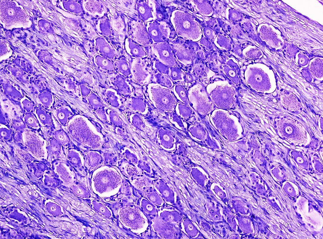
| Light microscopy of a spinal sensory ganglion also known as a dorsal root ganglion. The ganglion is formed of a cluster of nerve cell bodies each with a central nucleus and densely stained cytoplasm (purple). Between the cell bodies are many myelinated axons that convey sensory signals from peripheral nerves to the spinal cord via the spinal ganglia. Magnification x100 when narrow width printed at 10 cm | |
| Licence : | Droits gérés |
| Crédit: | Science Photo Library / Microscape |
| Taille de l’image : | 4884 px × 3613 px |
| Model Release : | Non requis |
| Property Release : | Non requis |
| Restrictions : | - |
Prix pour cette image À partir de 45 €
Produit vendu
(Calendrier, Carte postale, Carte de vœux, Impression sur textile, Packaging etc)
À partir de 45 €
Usage commercial
(Affichage, Annonce presse, Annonce TV, Carte, Digital - hors rés. sociaux, Digital - rés. sociaux etc)
À partir de 45 €
Éditorial
(Digital, Journal, Livre, Livre pratique, Magazine, Télévision etc)
À partir de 60 €
Usage non-commercial
(Digital - hors rés. sociaux, Digital - rés. sociaux etc)
À partir de 120 €