Motor neurons,light micrograph
Numéro d’image : 11704738
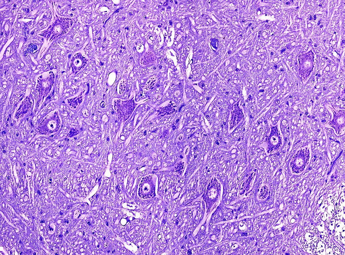
| Light microscopy of motor neurons in spinal cord grey matter. The cell bodies of motor neurons show a central nucleus and peripheral cytoplasm (deep purple). In histologic sections processes of dendrites and an axon extend from each cell body. In humans one such axon may be a metre in length if supplying the foot. Tissue between the cell bodies is called the neuropil and is formed of intermingling dendrites,axons,capillaries and glial cells. Magnification x100 when narrow width printed at 10 cm | |
| Licence : | Droits gérés |
| Crédit: | Science Photo Library / Microscape |
| Taille de l’image : | 4884 px × 3619 px |
| Model Release : | Non requis |
| Property Release : | Non requis |
| Restrictions : | - |
Prix pour cette image À partir de 45 €
Produit vendu
(Calendrier, Carte postale, Carte de vœux, Impression sur textile, Packaging etc)
À partir de 45 €
Usage commercial
(Affichage, Annonce presse, Annonce TV, Carte, Digital - hors rés. sociaux, Digital - rés. sociaux etc)
À partir de 45 €
Éditorial
(Digital, Journal, Livre, Livre pratique, Magazine, Télévision etc)
À partir de 60 €
Usage non-commercial
(Digital - hors rés. sociaux, Digital - rés. sociaux etc)
À partir de 120 €