Articular cartilage,light micrograph
Numéro d’image : 11704732
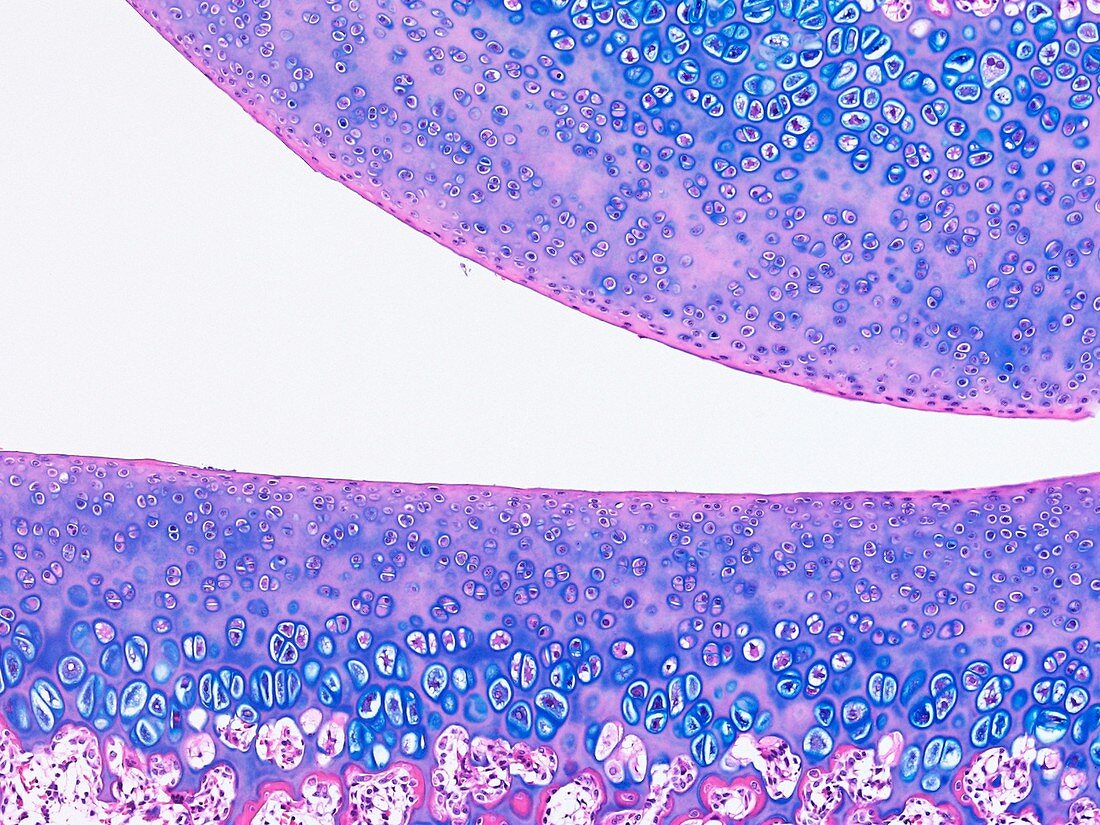
| Light microscopy of cartilage covering the surfaces of two bone ends inside a joint. Called hyaline articular cartilage it consists of a matrix and cartilage cells (chondrocytes) stained blue. Some functions of this cartilage are to provide an almost frictionless surface for joint movement and,by retaining water in its matrix,to resist compression. The joint space is filled with synovial fluid acting as a lubricant and nutrient source. The cartilage has no direct blood vessels. Magnification x120 when narrow width printed at 10 cm | |
| Licence : | Droits gérés |
| Crédit: | Science Photo Library / Microscape |
| Taille de l’image : | 4859 px × 3644 px |
| Model Release : | Non requis |
| Property Release : | Non requis |
| Restrictions : | - |
Prix pour cette image À partir de 45 €
Produit vendu
(Calendrier, Carte postale, Carte de vœux, Impression sur textile, Packaging etc)
À partir de 45 €
Usage commercial
(Affichage, Annonce presse, Annonce TV, Carte, Digital - hors rés. sociaux, Digital - rés. sociaux etc)
À partir de 45 €
Éditorial
(Digital, Journal, Livre, Livre pratique, Magazine, Télévision etc)
À partir de 60 €
Usage non-commercial
(Digital - hors rés. sociaux, Digital - rés. sociaux etc)
À partir de 120 €