Intervertebral disc,light micrograph
Numéro d’image : 11704726
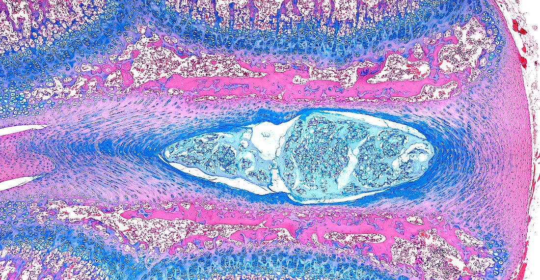
| Light microscopy of an intervertebral disc. The disc is found between two vertebral bodies their bone tissue margins stained pink. The centre of the disc is formed of cells and a gel-like matrix,called the nucleus pulposus. It is a remnant of the head-to-tail axis of the early embryo (the notochord). Fibrocartilage rings (blue) form the outer margins of the disc,called the nucleus fibrosus. The disc acts like a cushion between the stacked vertebral bodies and resists compression but allows movements of each vertebra. A slipped disc' is an abnormal protrusion (herniation) of the central portion through a damaged region or tear of the fibrocartilage. Magnification x60 when narrow width printed at 10 cm | |
| Licence : | Droits gérés |
| Crédit: | Science Photo Library / Microscape |
| Taille de l’image : | 5851 px × 3034 px |
| Model Release : | Non requis |
| Property Release : | Non requis |
| Restrictions : | - |
Prix pour cette image À partir de 45 €
Produit vendu
(Calendrier, Carte postale, Carte de vœux, Impression sur textile, Packaging etc)
À partir de 45 €
Usage commercial
(Affichage, Annonce presse, Annonce TV, Carte, Digital - hors rés. sociaux, Digital - rés. sociaux etc)
À partir de 45 €
Éditorial
(Digital, Journal, Livre, Livre pratique, Magazine, Télévision etc)
À partir de 60 €
Usage non-commercial
(Digital - hors rés. sociaux, Digital - rés. sociaux etc)
À partir de 120 €
Mots clés
- anneau de cartilage fibreux,
- annulus fibrosus,
- biologie,
- biologique,
- cartilage,
- catégorie,
- chondrocytes,
- colonne vertébrale,
- coupe,
- croissance osseuse,
- disque intervertébral,
- histologie,
- histologique,
- matrice du cartilage,
- microscope optique,
- microscopie optique,
- noyau gélatineux,
- noyau pulpeux,
- nucleus pulposus,
- os,
- partie,
- section,
- vertebra,
- vertebrae,
- vertèbre,
- vertèbres