Bone joint space,light micrograph
Numéro d’image : 11704724
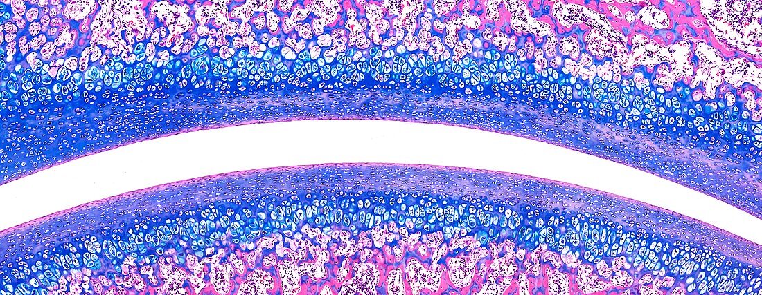
| Light microscopy of tissues covering the surfaces of two bone ends inside a joint. The opposing bone surfaces are covered with hyaline cartilage consisting of a matrix and cartilage cells (chondrocytes) stained blue. Beneath the cartilage zone is spongy bone (pink and blue) with gaps for bone marrow. The joint space is filled with synovial fluid acting as a lubricant and nutrient source. The cartilage has no direct blood vessels. Magnification x80 when narrow width printed at 10 cm | |
| Licence : | Droits gérés |
| Crédit: | Science Photo Library / Microscape |
| Taille de l’image : | 6732 px × 2616 px |
| Model Release : | Non requis |
| Property Release : | Non requis |
| Restrictions : | - |
Prix pour cette image À partir de 45 €
Produit vendu
(Calendrier, Carte postale, Carte de vœux, Impression sur textile, Packaging etc)
À partir de 45 €
Usage commercial
(Affichage, Annonce presse, Annonce TV, Carte, Digital - hors rés. sociaux, Digital - rés. sociaux etc)
À partir de 45 €
Éditorial
(Digital, Journal, Livre, Livre pratique, Magazine, Télévision etc)
À partir de 60 €
Usage non-commercial
(Digital - hors rés. sociaux, Digital - rés. sociaux etc)
À partir de 120 €