Fetal tooth,light micrograph
Numéro d’image : 11704322
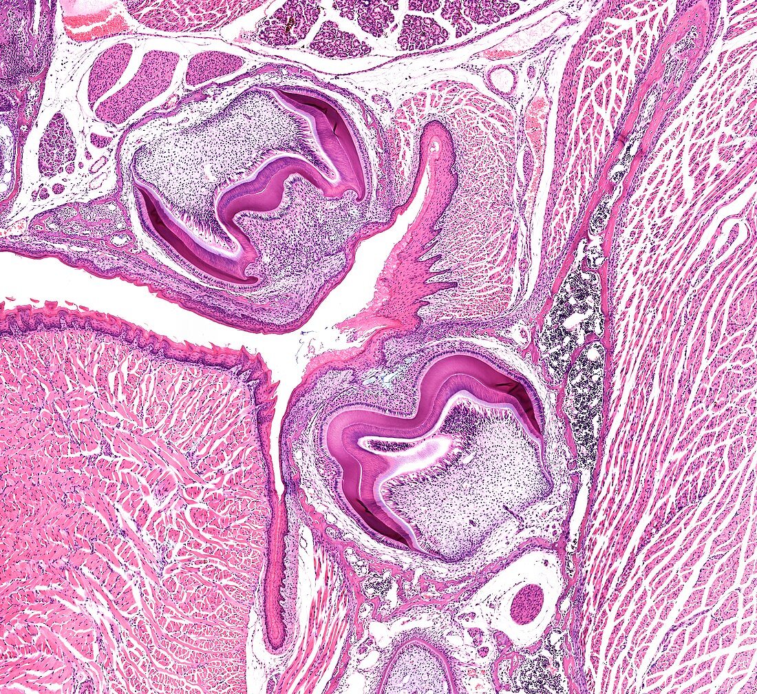
| Light microscopy of fetal teeth in an advanced stage of development. Each tooth is located below the gumline in a socket formed in the bone of the jaw. Part of the tongue is to the left. The growing crown (magenta curvatures) of the teeth is well defined,and show a sculptured appearance suggestive of the structure of molar teeth. The outermost layer is the growing enamel next to which is the dentin. The core of each tooth will become the pulp or root cavity where nerves and blood vessels supply the tooth. The region between the tooth and the jawbone will form the periodontal ligament that anchors a tooth in the socket-type recess. Magnification x40 when printed at 10 cm | |
| Licence : | Droits gérés |
| Crédit: | Science Photo Library / Microscape |
| Taille de l’image : | 4366 px × 4002 px |
| Model Release : | Non requis |
| Property Release : | Non requis |
| Restrictions : | - |
Prix pour cette image À partir de 45 €
Produit vendu
(Calendrier, Carte postale, Carte de vœux, Impression sur textile, Packaging etc)
À partir de 45 €
Usage commercial
(Affichage, Annonce presse, Annonce TV, Carte, Digital - hors rés. sociaux, Digital - rés. sociaux etc)
À partir de 45 €
Éditorial
(Digital, Journal, Livre, Livre pratique, Magazine, Télévision etc)
À partir de 60 €
Usage non-commercial
(Digital - hors rés. sociaux, Digital - rés. sociaux etc)
À partir de 120 €