Fetal eye,light micrograph
Numéro d’image : 11704300
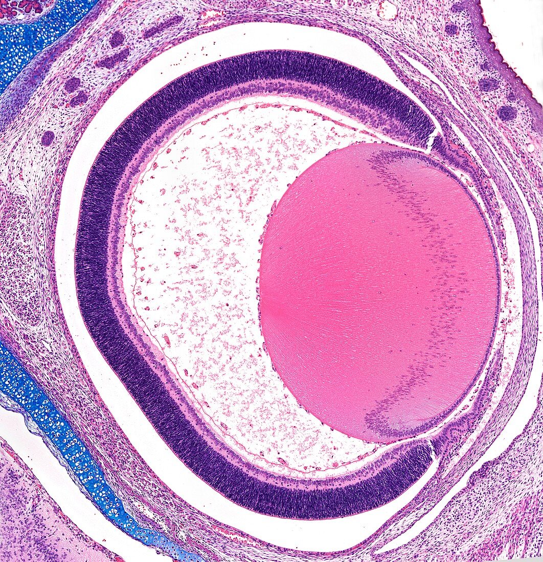
| Light micrograph of a section through an eye from a developing fetus. From right to left,this view shows the cornea,part of the iris,the developing lens (rounded area left of centre),the retina (upper and lower centre),and the cavity between the lens and retina,the vitreous body. The developing lens has a surface population of cells that progressively multiply and elongate into long lens fibres that make up the substance of the lens. Magnification x50 when printed at 10 cm | |
| Licence : | Droits gérés |
| Crédit: | Science Photo Library / Microscape |
| Taille de l’image : | 4134 px × 4282 px |
| Model Release : | Non requis |
| Property Release : | Non requis |
| Restrictions : | - |
Prix pour cette image À partir de 45 €
Produit vendu
(Calendrier, Carte postale, Carte de vœux, Impression sur textile, Packaging etc)
À partir de 45 €
Usage commercial
(Affichage, Annonce presse, Annonce TV, Carte, Digital - hors rés. sociaux, Digital - rés. sociaux etc)
À partir de 45 €
Éditorial
(Digital, Journal, Livre, Livre pratique, Magazine, Télévision etc)
À partir de 60 €
Usage non-commercial
(Digital - hors rés. sociaux, Digital - rés. sociaux etc)
À partir de 120 €