Lens of eye,light micrograph
Numéro d’image : 11704286
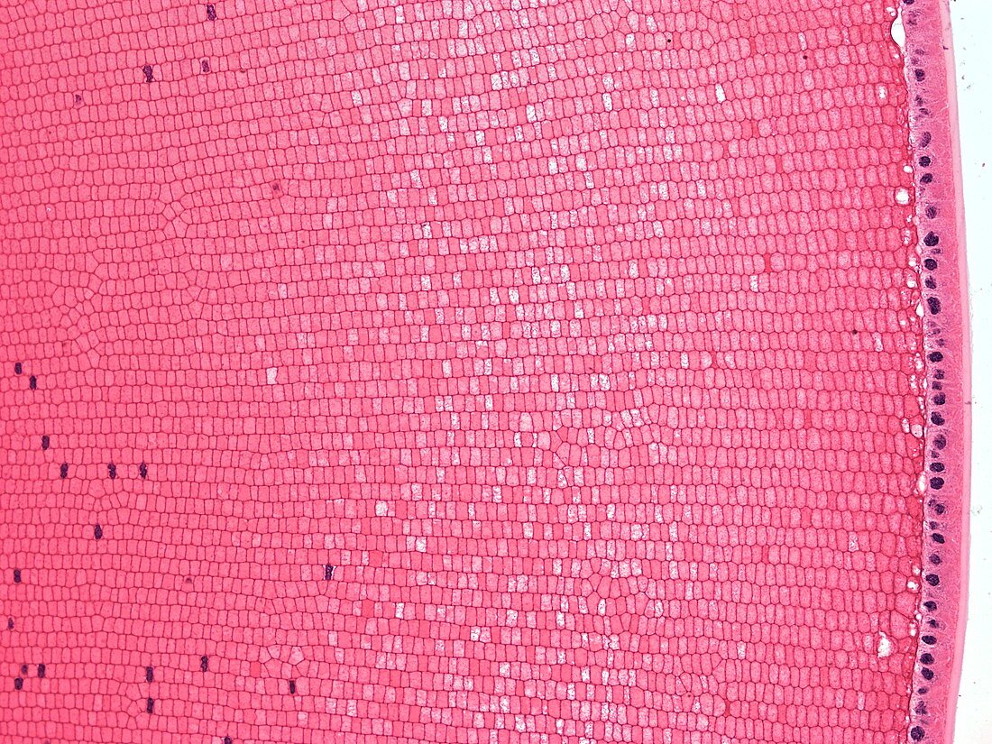
| Light microscopy of fibres in the lens of the eye. The lens is covered on its front surface by a capsule and epithelial cells. At the sides of the lens,the epithelial cells gradually elongate and then lose their nuclei,forming many thousands of long,thin tightly packed fibres seen here in cross-section. Each fibre contains a high abundance of transparent proteins called crystallins. Occasional fibres show a nucleus but eventually these disappear during lens development. The earliest lens fibres made in fetal life lie in the centre of the lens where they are retained. New lens fibres are added to the inner layers and may be formed throughout life. Magnification x180 when printed at 10 cm | |
| Licence : | Droits gérés |
| Crédit: | Science Photo Library / Microscape |
| Taille de l’image : | 4827 px × 3620 px |
| Model Release : | Non requis |
| Property Release : | Non requis |
| Restrictions : | - |
Prix pour cette image À partir de 45 €
Produit vendu
(Calendrier, Carte postale, Carte de vœux, Impression sur textile, Packaging etc)
À partir de 45 €
Usage commercial
(Affichage, Annonce presse, Annonce TV, Carte, Digital - hors rés. sociaux, Digital - rés. sociaux etc)
À partir de 45 €
Éditorial
(Digital, Journal, Livre, Livre pratique, Magazine, Télévision etc)
À partir de 60 €
Usage non-commercial
(Digital - hors rés. sociaux, Digital - rés. sociaux etc)
À partir de 120 €