Intestinal lymphoid tissue
Numéro d’image : 11704282
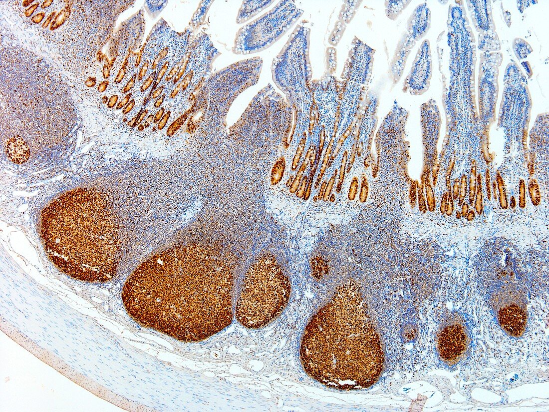
| Light microscopy of lymphoid follicles in a Peyer's patch of the ileum,the last segment of the small bowel. The lymphoid cells are stained brown with a immunostain for any cells that are about to divide or are proliferating. Dividing cells in the intestinal crypts are similarly stained. The follicles supply lymphocytes to the local tissue for immune defence against harmful antigens such as toxins and infectious micro-organisms. Peyer's patches and lymphoid components of the intestines are collectively referred to as gut-associated lymphoid tissue abbreviated as GALT. Magnification x40 when printed at 10 cm | |
| Licence : | Droits gérés |
| Crédit: | Science Photo Library / Microscape |
| Taille de l’image : | 4827 px × 3620 px |
| Model Release : | Non requis |
| Property Release : | Non requis |
| Restrictions : | - |
Prix pour cette image À partir de 45 €
Produit vendu
(Calendrier, Carte postale, Carte de vœux, Impression sur textile, Packaging etc)
À partir de 45 €
Usage commercial
(Affichage, Annonce presse, Annonce TV, Carte, Digital - hors rés. sociaux, Digital - rés. sociaux etc)
À partir de 45 €
Éditorial
(Digital, Journal, Livre, Livre pratique, Magazine, Télévision etc)
À partir de 60 €
Usage non-commercial
(Digital - hors rés. sociaux, Digital - rés. sociaux etc)
À partir de 120 €
Mots clés
- biologie,
- biologique,
- catégorie,
- coupe,
- en bonne santé,
- follicule lymphoïde,
- gut,
- histologie,
- histologique,
- iléon,
- ileum,
- intestin,
- intestin grêle,
- lymphocytes,
- microscope optique,
- microscope photonique,
- microscopie optique,
- microscopie photonique,
- normal,
- partie,
- petit intestin,
- sain,
- section,
- système immunitaire,
- tissu lymphoïde