Yeast cell nucleus,SIM micrograph
Numéro d’image : 11702765
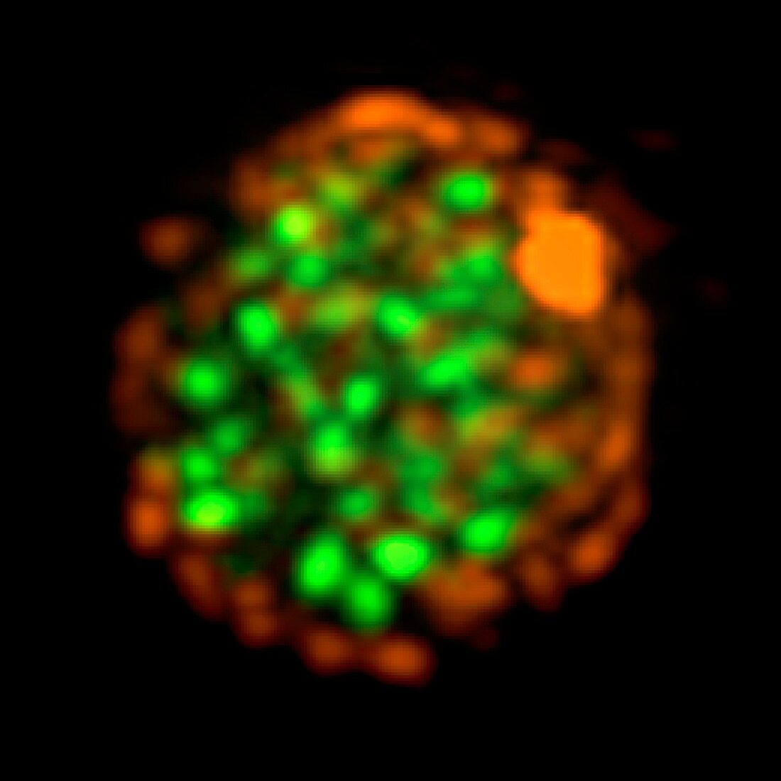
| Yeast cell nucleus. Fluorescent structured illumination micrograph (SIM) of replication factories within a yeast cell nucleus. The colours represent: proliferating cell nuclear antigen (PCNA) protein (green),Nic96 nucleoporin protein of the nuclear pore complex (orange),and the spindle pole protein spc42 (orange dot,upper right). This image was obtained using structured illumination microscopy,a technique that allows light microscopy to bypass the resolution limits imposed by the diffraction of light. This is OMX structured illumination microscopy,which uses 15 orientations and Fourier transform computer analysis | |
| Licence : | Droits gérés |
| Crédit: | Science Photo Library / DR PAUL ANDREWS, UNIVERSITY OF DUNDEE |
| Taille de l’image : | 3236 px × 3236 px |
| Model Release : | Non requis |
| Property Release : | Non requis |
| Restrictions : | - |
Prix pour cette image À partir de 45 €
Produit vendu
(Calendrier, Carte postale, Carte de vœux, Impression sur textile, Packaging etc)
À partir de 45 €
Usage commercial
(Affichage, Annonce presse, Annonce TV, Carte, Digital - hors rés. sociaux, Digital - rés. sociaux etc)
À partir de 45 €
Éditorial
(Digital, Journal, Livre, Livre pratique, Magazine, Télévision etc)
À partir de 60 €
Usage non-commercial
(Digital - hors rés. sociaux, Digital - rés. sociaux etc)
À partir de 120 €