Keratinocyte,SIM micrograph
Numéro d’image : 11702764
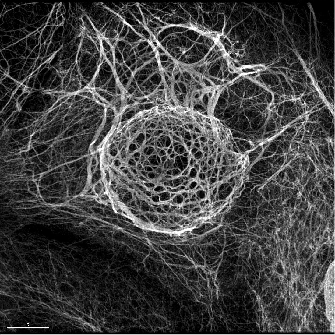
| Keratinocyte. Structured illumination micrograph (SIM) of the intermediate filament network of a keratinocyte,as visualised with an antibody directed against the keratin 14 protein. Keratinocytes are the major type of skin cell,changing as they pass through the skin and eventually becoming dead keratinised cells at the skin's surface. This image was obtained using structured illumination microscopy,a technique that allows light microscopy to bypass the resolution limits imposed by the diffraction of light. This is OMX structured illumination microscopy,which uses 15 orientations and Fourier transform computer analysis | |
| Licence : | Droits gérés |
| Crédit: | Science Photo Library / DR PAUL ANDREWS, UNIVERSITY OF DUNDEE |
| Taille de l’image : | 2965 px × 2965 px |
| Model Release : | Non requis |
| Property Release : | Non requis |
| Restrictions : | - |
Prix pour cette image À partir de 45 €
Produit vendu
(Calendrier, Carte postale, Carte de vœux, Impression sur textile, Packaging etc)
À partir de 45 €
Usage commercial
(Affichage, Annonce presse, Annonce TV, Carte, Digital - hors rés. sociaux, Digital - rés. sociaux etc)
À partir de 45 €
Éditorial
(Digital, Journal, Livre, Livre pratique, Magazine, Télévision etc)
À partir de 60 €
Usage non-commercial
(Digital - hors rés. sociaux, Digital - rés. sociaux etc)
À partir de 120 €
Mots clés
- aucun,
- biochimie,
- biochimique,
- biologie,
- biologie cellulaire,
- biologique,
- cellulaire,
- cellule,
- corps humain,
- cytologie,
- cytologique,
- dermatologie,
- dermatologique,
- filament,
- kératinocytes,
- micrographie optique,
- microscope optique,
- microscopie optique,
- monochrome,
- n/b,
- noir et blanc,
- noir-et-blanc,
- peau,
- personne,
- protéine