Snail,micro-CT scan
Numéro d’image : 11686786
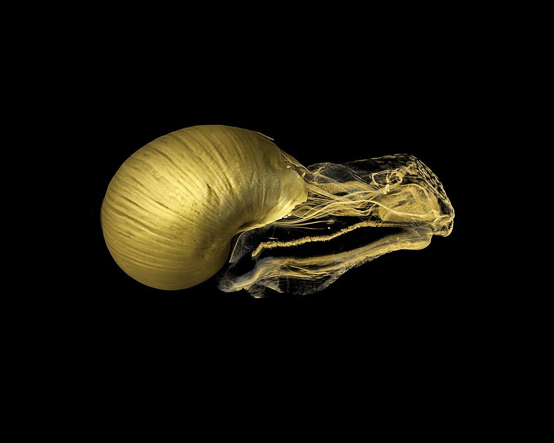
| Snail (Scutalus sp.),micro-CT scan. This image was acquired using X-ray micro-Computed Tomography (also known as micro-CT). A micro-CT scanner projects a beam of X-rays through the sample onto a detector panel. Images are collected over a 360° rotation and these are then reconstructed to form a virtual 3D model of the specimen. This virtual object that can be viewed from all angles and sliced open or digitally dissected. This specimen was stained with iodine to provide contrast in the soft tissue,allowing features such as the nerves and muscles to be visualised. Image by Dan Sykes | |
| Licence : | Droits gérés |
| Crédit: | Science Photo Library / NATURAL HISTORY MUSEUM, LONDON / DAN SYKES |
| Taille de l’image : | 3309 px × 2646 px |
| Model Release : | Non requis |
| Property Release : | Non requis |
| Restrictions : | - |
Prix pour cette image À partir de 45 €
Produit vendu
(Calendrier, Carte postale, Carte de vœux, Impression sur textile, Packaging etc)
À partir de 45 €
Usage commercial
(Affichage, Annonce presse, Annonce TV, Carte, Digital - hors rés. sociaux, Digital - rés. sociaux etc)
À partir de 45 €
Éditorial
(Digital, Journal, Livre, Livre pratique, Magazine, Télévision etc)
À partir de 60 €
Usage non-commercial
(Digital - hors rés. sociaux, Digital - rés. sociaux etc)
À partir de 120 €
Mots clés
- 3 dimensions,
- 3D,
- anatomie,
- animal,
- biologie,
- biologique,
- escargot,
- faune,
- machine à rayons X,
- METRIS X-TEK HMX ST 225,
- micro-CT,
- microtomographie aux rayons X,
- nature,
- nikon,
- radiographie,
- rayons X,
- représentation,
- scanner tomographique assisté par ordinateur ordinateur,
- scanner tomographique par mico-ordinateur,
- scanner tomographique par ordinateur,
- tomodensitométrie,
- tomographie assistée par ordinateur,
- zoologie,
- zoologique