Alzheimer's brain,computer artwork
Numéro d’image : 11685719
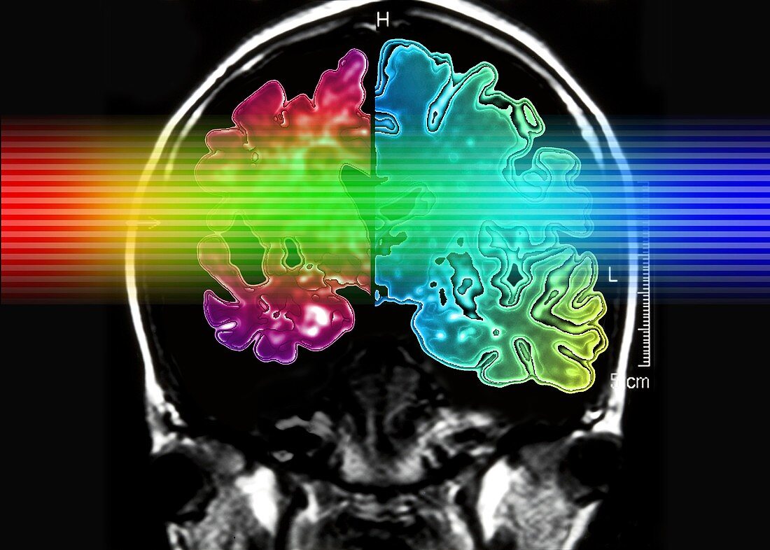
| Alzheimer's brain. Computer graphic of a vertical (coronal) slice through the brain of an Alzheimer patient (at left) compared with a normal brain (at right),overlaid a magnetic resonance image (MRI) of a human head. The colour gradient is depicting the MRI scanning process.The Alzheimer's disease brain (red) is considerably shrunken,due to the degeneration and death of nerve cells. Apart from a decrease in brain volume,the surface of the brain is often more deeply folded. Tangled protein filaments (neurofibrillary tangles) occur within nerve cells and patients also develop brain lesions of beta- amyloid protein. Alzheimer's disease accounts for most cases of senile dementia. Symptoms include memory loss,disorientation,personality change and delusion. It ultimately leads to death | |
| Licence : | Droits gérés |
| Crédit: | Science Photo Library / Pasieka, Alfred |
| Taille de l’image : | 5500 px × 3930 px |
| Model Release : | Non requis |
| Property Release : | Non requis |
| Restrictions : | - |
Prix pour cette image À partir de 45 €
Produit vendu
(Calendrier, Carte postale, Carte de vœux, Impression sur textile, Packaging etc)
À partir de 45 €
Usage commercial
(Affichage, Annonce presse, Annonce TV, Carte, Digital - hors rés. sociaux, Digital - rés. sociaux etc)
À partir de 45 €
Éditorial
(Digital, Journal, Livre, Livre pratique, Magazine, Télévision etc)
À partir de 60 €
Usage non-commercial
(Digital - hors rés. sociaux, Digital - rés. sociaux etc)
À partir de 120 €