Skull fracture surgery,artwork
Numéro d’image : 11681847
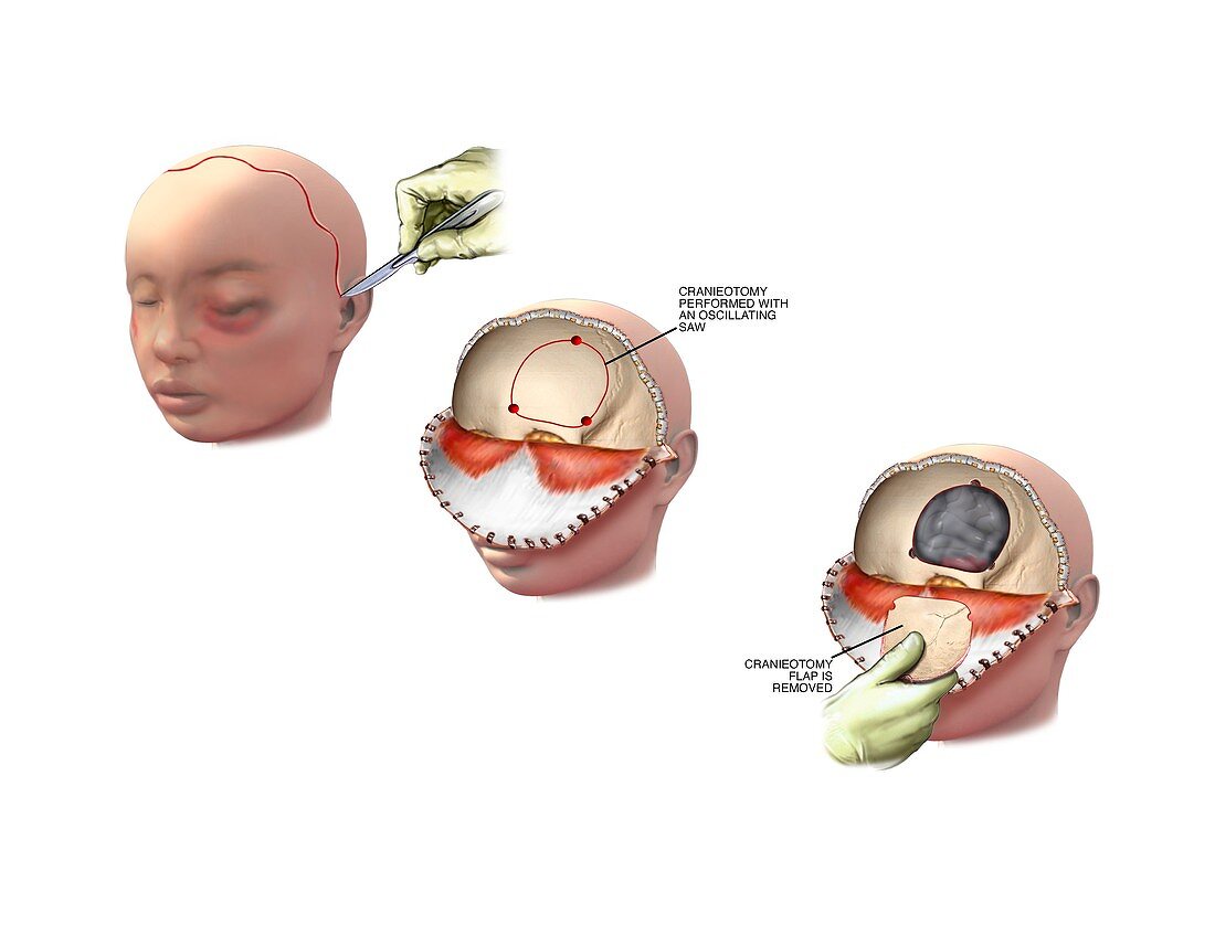
| Skull fracture surgery. Artwork sequence showing stages in surgery for open reduction and internal fixation of a fracture of the orbital part of the frontal bone of the skull. At upper left is the incision site,with a scalpel used by a surgeon. At centre,the scalp has been drawn forward (a bicoronal flap) to expose the superior orbital rim. An oscillating bone saw is used to perform a craniotomy where a section of the skull is removed to expose the brain (lower right). This may be done to relieve brain swelling and pressure caused by the blunt force trauma that fractured the skull. The skull flap may be repaired or replaced,secured with metal plates and screws | |
| Licence : | Droits gérés |
| Crédit: | Science Photo Library / Alesi, John T. |
| Taille de l’image : | 4805 px × 3661 px |
| Model Release : | Non requis |
| Property Release : | Non requis |
| Restrictions : | - |
Prix pour cette image À partir de 45 €
Produit vendu
(Calendrier, Carte postale, Carte de vœux, Impression sur textile, Packaging etc)
À partir de 45 €
Usage commercial
(Affichage, Annonce presse, Annonce TV, Carte, Digital - hors rés. sociaux, Digital - rés. sociaux etc)
À partir de 45 €
Éditorial
(Digital, Journal, Livre, Livre pratique, Magazine, Télévision etc)
À partir de 60 €
Usage non-commercial
(Digital - hors rés. sociaux, Digital - rés. sociaux etc)
À partir de 120 €
Mots clés
- aucun,
- cassé,
- chirurgical,
- coincé,
- corps humain,
- crâne,
- épinglé,
- fixation,
- fixée,
- fracture,
- fracturé,
- illustration,
- implant,
- implants métal,
- implants métalliques,
- médecine,
- médical,
- médicale,
- oeuvre,
- opération chirurgicale,
- os,
- os cassé,
- ostéologie,
- personne,
- plaque,
- schéma,
- séquence,
- séries,
- soins de santé,
- technologie,
- technologique,
- traitement