Multiple facial skull fractures
Numéro d’image : 11681843
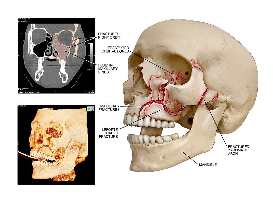
| Multiple facial skull fractures. Labelled artwork and scans of multiple fractures of the facial bones of the left side of the skull. At upper left is a coronal computed tomography (CT) scan showing fluid producing opacification (pink) in the maxillary sinus,as well as showing the broken bones. At lower left is a 3D model that is a compilation of scans made to assess the extent of the injuries. The main artwork is an anterolateral view showing fractures of the orbital bones of the left eye,fractures of the maxilla bone,a Leforte grade I fracture,and fractures of the left zygomatic arch. These fractures would all typically be caused by blunt force trauma and require surgical intervention | |
| Licence : | Droits gérés |
| Crédit: | Science Photo Library / Alesi, John T. |
| Taille de l’image : | 4860 px × 3614 px |
| Model Release : | Non requis |
| Property Release : | Non requis |
| Restrictions : | - |
Prix pour cette image À partir de 45 €
Produit vendu
(Calendrier, Carte postale, Carte de vœux, Impression sur textile, Packaging etc)
À partir de 45 €
Usage commercial
(Affichage, Annonce presse, Annonce TV, Carte, Digital - hors rés. sociaux, Digital - rés. sociaux etc)
À partir de 45 €
Éditorial
(Digital, Journal, Livre, Livre pratique, Magazine, Télévision etc)
À partir de 60 €
Usage non-commercial
(Digital - hors rés. sociaux, Digital - rés. sociaux etc)
À partir de 120 €