Healthy knee,CT scan
Numéro d’image : 11659024
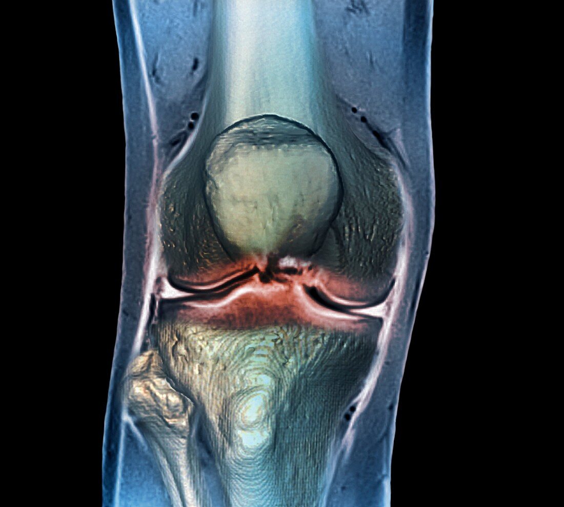
| Healthy knee. Coloured frontal computed tomography (CT) scan projected on to a magnetic resonance imaging (MRI) scan of the knee of a 30 year old. The knee joint is formed by the articulation of the femur (thigh bone,top) with tibia (shin bone,bottom centre). The smaller fibula (bottom left) is also seen. The patella (knee cap) is seen over the bottom of the femur | |
| Licence : | Droits gérés |
| Crédit: | Science Photo Library / Zephyr |
| Taille de l’image : | 4425 px × 3980 px |
| Model Release : | Non requis |
| Property Release : | Non requis |
| Restrictions : | - |
Prix pour cette image À partir de 45 €
Produit vendu
(Calendrier, Carte postale, Carte de vœux, Impression sur textile, Packaging etc)
À partir de 45 €
Usage commercial
(Affichage, Annonce presse, Annonce TV, Carte, Digital - hors rés. sociaux, Digital - rés. sociaux etc)
À partir de 45 €
Éditorial
(Digital, Journal, Livre, Livre pratique, Magazine, Télévision etc)
À partir de 60 €
Usage non-commercial
(Digital - hors rés. sociaux, Digital - rés. sociaux etc)
À partir de 120 €
Mots clés
- 30,
- adulte,
- anatomie,
- anatomique,
- années 30,
- articulation,
- avant,
- biologie,
- biologique,
- coloré,
- colorié,
- colorisé,
- composite,
- corps humain,
- devant,
- en bonne santé,
- fémur,
- fibula,
- frontal,
- genou,
- gens,
- I.R.M.,
- imagerie par résonnance magnétique,
- IRM,
- jambe,
- machine à rayons X,
- membre inférieur,
- normal,
- os,
- os de la cuisse,
- patella,
- péroné,
- personne,
- radiographie,
- rayons X,
- rotule,
- sain,
- scanner,
- tibia,
- tomodensitométrie,
- trente