Muscle contraction,artwork
Numéro d’image : 11597649
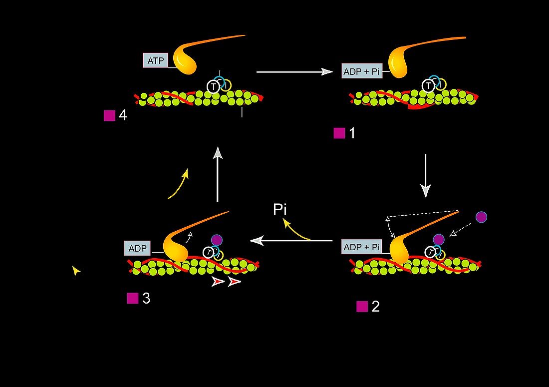
| Muscle contraction. Artwork showing the mechanism for muscle contraction in skeletal and cardiac muscle. At top right troponin (T,C,I) is bound to tropomyosin (red),preventing the myosin head (gold) from forming a crossbridge with the actin filament (green). The propagation of an action potential releases calcium ions (purple) into the muscle cell (bottom right). The calcium ion binds to troponin,changing its shape and exposing the myosin binding site on the actin filament. The binding of actin and mysoin causes the muscle to contract (bottom left). The myosin then binds ATP,which allows it to release the actin and return to the starting position (top left) | |
| Licence : | Droits gérés |
| Crédit: | Science Photo Library / Leroy, Francis / Biocosmos |
| Taille de l’image : | 5000 px × 3524 px |
| Model Release : | Non requis |
| Property Release : | Non requis |
| Restrictions : | - |
Prix pour cette image À partir de 45 €
Produit vendu
(Calendrier, Carte postale, Carte de vœux, Impression sur textile, Packaging etc)
À partir de 45 €
Usage commercial
(Affichage, Annonce presse, Annonce TV, Carte, Digital - hors rés. sociaux, Digital - rés. sociaux etc)
À partir de 45 €
Éditorial
(Digital, Journal, Livre, Livre pratique, Magazine, Télévision etc)
À partir de 60 €
Usage non-commercial
(Digital - hors rés. sociaux, Digital - rés. sociaux etc)
À partir de 120 €
Mots clés
- adjudication,
- anatomie,
- anatomique,
- arrière plan noir,
- arrière-plan noir,
- biologie,
- biologique,
- cardiaque,
- cellule musculaire,
- contracter,
- contraction musculaire,
- filaments d'actine,
- fond noir,
- glisser,
- humain,
- illustration,
- mécanisme,
- modèle,
- oeuvre,
- passation,
- physiologie,
- physiologique,
- protéine,
- squelettique,
- tension,
- tropomyosine,
- troponine