Brain tumour,MRI scan
Numéro d’image : 11572163
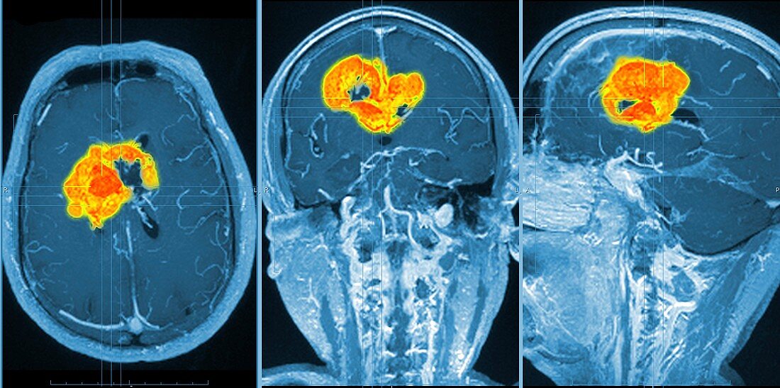
| Brain tumour. Coloured orthogonal views (axial left,coronal middle,sagittal right) of a male head,made from magnetic resonance imaging (MRI) scans. The scans reveal a large glioma tumour (yellow-orange),a central nervous system tumour that arises from glial cells. About half of all primary brain tumours are gliomas. MRI uses radio waves and a magnet to obtain "slice" body images | |
| Licence : | Droits gérés |
| Crédit: | Science Photo Library / Pasieka, Alfred |
| Taille de l’image : | 5928 px × 2948 px |
| Model Release : | Non requis |
| Property Release : | Non requis |
| Restrictions : | - |
Prix pour cette image À partir de 45 €
Produit vendu
(Calendrier, Carte postale, Carte de vœux, Impression sur textile, Packaging etc)
À partir de 45 €
Usage commercial
(Affichage, Annonce presse, Annonce TV, Carte, Digital - hors rés. sociaux, Digital - rés. sociaux etc)
À partir de 45 €
Éditorial
(Digital, Journal, Livre, Livre pratique, Magazine, Télévision etc)
À partir de 60 €
Usage non-commercial
(Digital - hors rés. sociaux, Digital - rés. sociaux etc)
À partir de 120 €
Mots clés
- 3 D,
- 3 dimensions,
- 3-D,
- 3D,
- central,
- cérébral,
- cerveau,
- coloré,
- colorié,
- colorisé,
- corps,
- coupe,
- coupé,
- couper,
- découpe,
- découpé,
- diagnostic,
- diagnostique,
- dimensionnel,
- glioma,
- gliome,
- humain,
- loin,
- magnétique,
- maladie,
- nerveux,
- neuro-imagerie,
- neuroimagerie,
- personne,
- plan en coupe,
- représentation,
- résonance,
- S.N.C.,
- scanner,
- SNC,
- système,
- système nerveux central,
- tête,
- tridimensionnel,
- trois dimensions