Parkinson's disease brain,MRI scan
Numéro d’image : 11567738
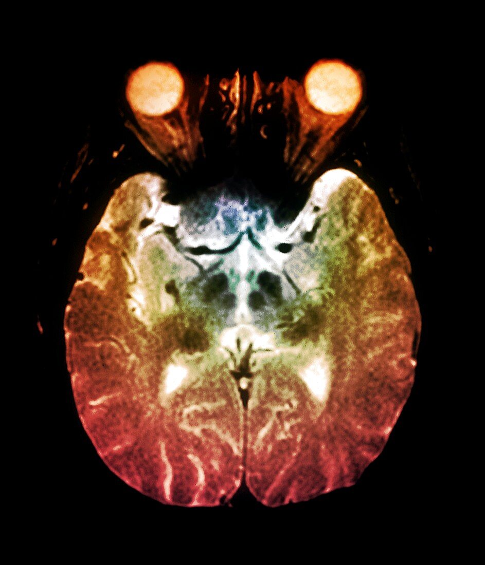
| Coloured Magnetic Resonance Imaging (MRI) scan in axial section through the brain of a 65 year old patient,showing Parkinson's disease. At centre are a number of plaques (dark areas) in the thalamus close to the basal ganglia. Parkinson's disease is caused by loss of nerve cells in the basal ganglia resulting in jerky,involuntary movements | |
| Licence : | Droits gérés |
| Crédit: | Science Photo Library / Zephyr |
| Taille de l’image : | 3886 px × 4533 px |
| Model Release : | Non requis |
| Property Release : | Non requis |
| Restrictions : | - |
Prix pour cette image À partir de 45 €
Produit vendu
(Calendrier, Carte postale, Carte de vœux, Impression sur textile, Packaging etc)
À partir de 45 €
Usage commercial
(Affichage, Annonce presse, Annonce TV, Carte, Digital - hors rés. sociaux, Digital - rés. sociaux etc)
À partir de 45 €
Éditorial
(Digital, Journal, Livre, Livre pratique, Magazine, Télévision etc)
À partir de 60 €
Usage non-commercial
(Digital - hors rés. sociaux, Digital - rés. sociaux etc)
À partir de 120 €
Mots clés
- cerveau,
- coupe longitudinale,
- désordre,
- diagnostic,
- diagnostique,
- état,
- I.R.M.,
- imagerie par résonance magnétique,
- imagerie par résonnance magnétique,
- IRM,
- maladie,
- maladie de Parkinson,
- médical,
- médicale,
- neuro-imagerie,
- neuroimagerie,
- noyaux gris centraux,
- parkinson,
- personnes âgées,
- plaque,
- plaques,
- soins de santé,
- thalamus,
- trouble