Confocal microscope,artwork
Numéro d’image : 11563677
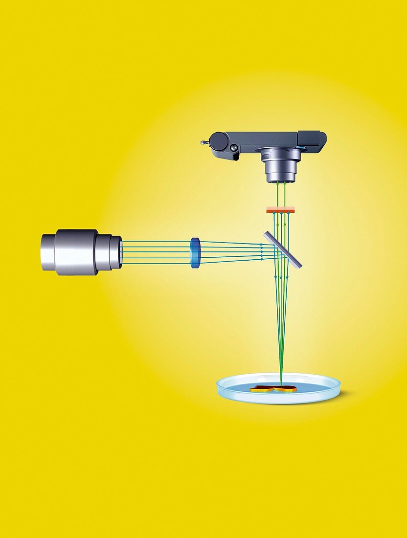
| Confocal light microscope,artwork. Confocal microscopy uses fluorescent dyes to highlight tissues,cellular structures and proteins in the samples being analysed. A laser beam (blue lines,source at left) is passed through a lens (blue oval) that focuses it on to a beam splitter (grey diagonal line). Some of the laser beams are directed to the sample (bottom right). The fluorescent dyes in the sample are excited by the laser beams and emit fluorescent light. This light passes back through the beam splitter and is collected by a digital camera. Laser beams directed towards the camera are removed by a filter (orange horizontal line) | |
| Licence : | Droits gérés |
| Crédit: | Science Photo Library / Lunau, Claus |
| Taille de l’image : | 3664 px × 4843 px |
| Model Release : | Non requis |
| Property Release : | Non requis |
| Restrictions : | - |
Prix pour cette image À partir de 45 €
Produit vendu
(Calendrier, Carte postale, Carte de vœux, Impression sur textile, Packaging etc)
À partir de 45 €
Usage commercial
(Affichage, Annonce presse, Annonce TV, Carte, Digital - hors rés. sociaux, Digital - rés. sociaux etc)
À partir de 45 €
Éditorial
(Digital, Journal, Livre, Livre pratique, Magazine, Télévision etc)
À partir de 60 €
Usage non-commercial
(Digital - hors rés. sociaux, Digital - rés. sociaux etc)
À partir de 120 €
Mots clés
- agrandissement,
- appareil photo digital,
- appareil photo numérique,
- arrière plan jaune,
- arrière-plan jaune,
- échantillon,
- équipement,
- escpèce,
- filtrer,
- fluorescence,
- fluorescent,
- fond jaune,
- illustration,
- laser,
- lentille,
- lumière fluorescente,
- machine,
- matériel,
- microscope confocal,
- miroir,
- oeuvre,
- physique,
- poutre,
- schéma,
- spécimen,
- technologie,
- technologique