Hip joint replacement (image 2 of 2)
Numéro d’image : 11562464
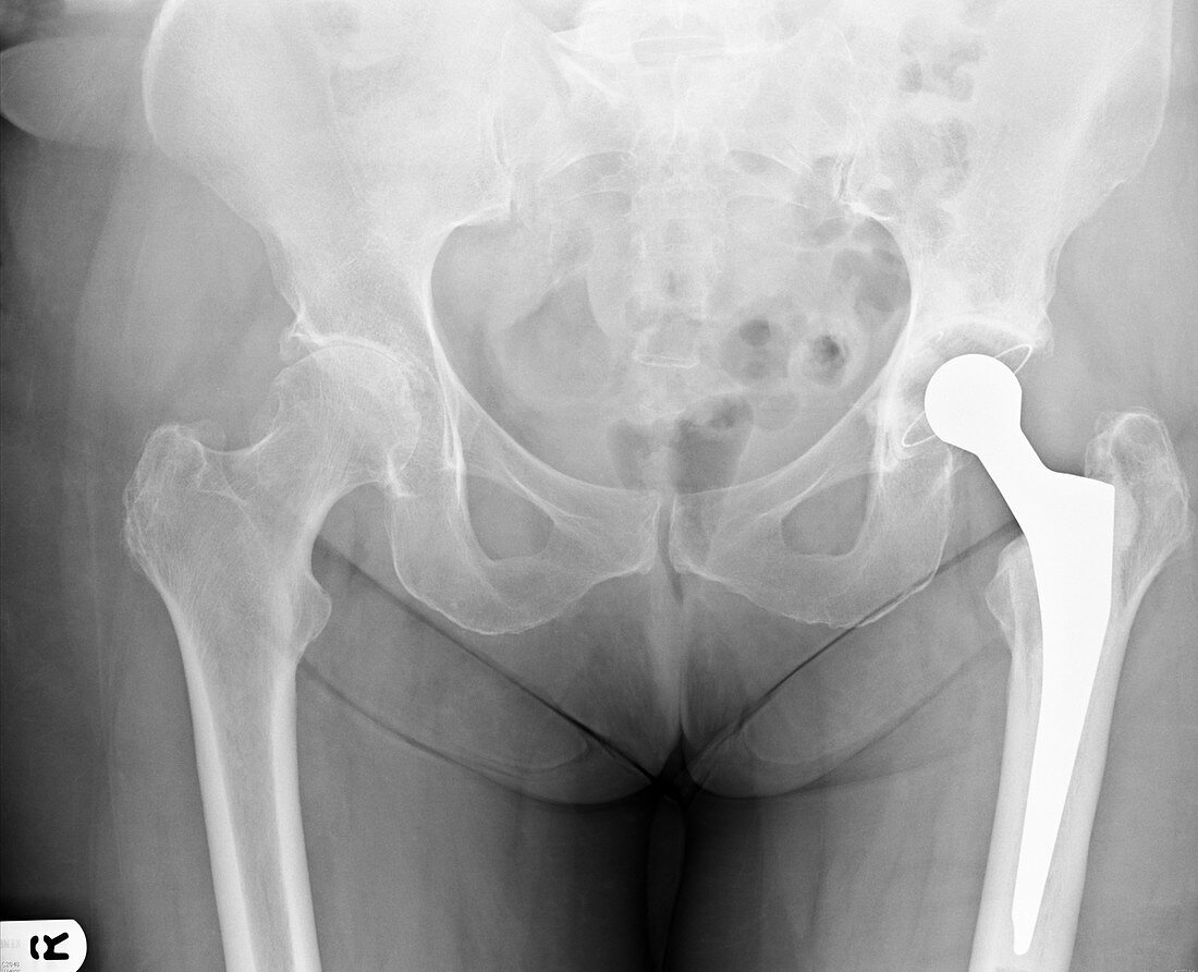
| X-ray (rear view) of the pelvis of an adult woman,showing an artificial (prosthetic) hip joint replacement. At right is a metal ball-and-socket implant forming a prosthetic joint between the pelvis and the thigh-bone (femur). The other hip joint (at left) reveals osteoarthritis with loss of space between the joint. See image 1 of this patient before surgery | |
| Licence : | Droits gérés |
| Crédit: | Science Photo Library / Marazzi, Dr. P. |
| Taille de l’image : | 4661 px × 3780 px |
| Model Release : | Non requis |
| Property Release : | Non requis |
| Restrictions : | - |
Prix pour cette image À partir de 45 €
Produit vendu
(Calendrier, Carte postale, Carte de vœux, Impression sur textile, Packaging etc)
À partir de 45 €
Usage commercial
(Affichage, Annonce presse, Annonce TV, Carte, Digital - hors rés. sociaux, Digital - rés. sociaux etc)
À partir de 45 €
Éditorial
(Digital, Journal, Livre, Livre pratique, Magazine, Télévision etc)
À partir de 60 €
Usage non-commercial
(Digital - hors rés. sociaux, Digital - rés. sociaux etc)
À partir de 120 €
Mots clés
- arthrite,
- arthritique,
- arthritis,
- arthrose,
- artificiel,
- désordre,
- état,
- femme adulte,
- fémur,
- maladie,
- médical,
- médicale,
- osteoarthritis,
- patient,
- patients,
- pelvien,
- pelvienne,
- pelvis,
- prothèse,
- prothèse totale de hanche,
- prothétique,
- PTH,
- radiographie,
- rayons X,
- soins de santé,
- traitement,
- trouble,
- vue arrière