'Dislocated hip replacement,X-ray'
Numéro d’image : 11560761
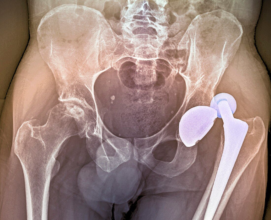
| Coloured X-ray (front view,supine) of the pelvis of a patient aged 36,showing dislocation of a total hip replacement. In this artificial (prosthetic) hip joint,the shaft and ball attached to the thigh bone (lower right) has dislocated from the socket fitted into the pelvic bone | |
| Licence : | Droits gérés |
| Crédit: | Science Photo Library / Zephyr |
| Taille de l’image : | 4654 px × 3784 px |
| Model Release : | Non requis |
| Property Release : | Non requis |
| Restrictions : | - |
Prix pour cette image À partir de 45 €
Produit vendu
(Calendrier, Carte postale, Carte de vœux, Impression sur textile, Packaging etc)
À partir de 45 €
Usage commercial
(Affichage, Annonce presse, Annonce TV, Carte, Digital - hors rés. sociaux, Digital - rés. sociaux etc)
À partir de 45 €
Éditorial
(Digital, Journal, Livre, Livre pratique, Magazine, Télévision etc)
À partir de 60 €
Usage non-commercial
(Digital - hors rés. sociaux, Digital - rés. sociaux etc)
À partir de 120 €
Mots clés
- adulte,
- âgé de,
- arthrite,
- arthritis,
- artificiel,
- désordre,
- disloqué,
- état,
- fémur,
- joint prosthétique,
- luxation,
- maladie,
- médical,
- médicale,
- os de la cuisse,
- pelvien,
- pelvienne,
- pelvis,
- prothèse,
- radiographie,
- rayon X coloré,
- rayons X,
- remplacement total de la hanche,
- soins de santé,
- traitement,
- trouble,
- vue de face,
- vue frontale