Double hip replacement,X-ray
Numéro d’image : 11560746
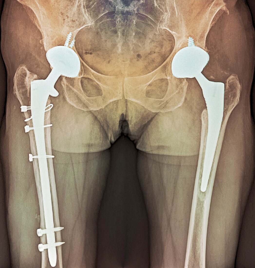
| Coloured X-ray (front view) of the pelvis of a patient aged 63,showing two total hip replacements. Each hip has been fitted with an artificial (prosthetic) socket and each thigh bone (femur) has been fitted with a ball and shaft,to form two new articulating joints. The prostheses differ in size and the method by which they have been fixed into the bone in this patient | |
| Licence : | Droits gérés |
| Crédit: | Science Photo Library / Zephyr |
| Taille de l’image : | 4090 px × 4299 px |
| Model Release : | Non requis |
| Property Release : | Non requis |
| Restrictions : | - |
Prix pour cette image À partir de 45 €
Produit vendu
(Calendrier, Carte postale, Carte de vœux, Impression sur textile, Packaging etc)
À partir de 45 €
Usage commercial
(Affichage, Annonce presse, Annonce TV, Carte, Digital - hors rés. sociaux, Digital - rés. sociaux etc)
À partir de 45 €
Éditorial
(Digital, Journal, Livre, Livre pratique, Magazine, Télévision etc)
À partir de 60 €
Usage non-commercial
(Digital - hors rés. sociaux, Digital - rés. sociaux etc)
À partir de 120 €
Mots clés
- adulte,
- âgé de,
- artificiel,
- désordre,
- deux,
- double,
- emboîtement de prothèse,
- fémur,
- maladie,
- médical,
- médicale,
- os de la cuisse,
- pelvien,
- pelvienne,
- pelvis,
- prothèse,
- radiographie,
- rayon X coloré,
- rayons X,
- remplacement total de la hanche,
- soins de santé,
- taille,
- traitement,
- trouble,
- vue de face,
- vue frontale