'Herniated spinal disc,MRI scan'
Numéro d’image : 11560742
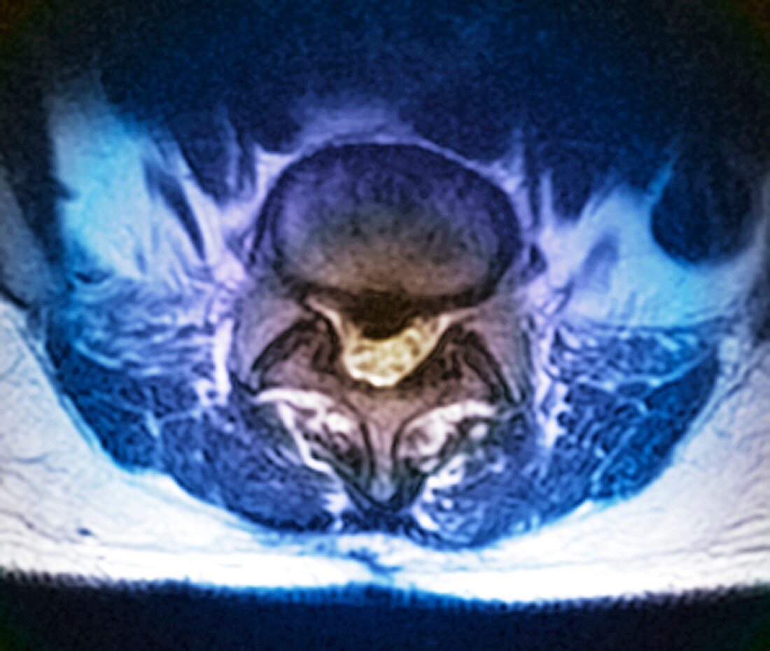
| Coloured magnetic resonance imaging (MRI) scan of an axial (horizontal) view of the spine of a patient aged 35,showing a herniated disc at upper centre (rounded) which has ruptured causing it to press on the spinal cord below (yellow). This disc is between lumbar vertebra (L5) and sacral vertebra (S1) in the lower back. Surgery was required to replace this damaged disc with an artificial (prosthetic) one | |
| Licence : | Droits gérés |
| Crédit: | Science Photo Library / Zephyr |
| Taille de l’image : | 4583 px × 3874 px |
| Model Release : | Non requis |
| Property Release : | Non requis |
| Restrictions : | - |
Prix pour cette image À partir de 45 €
Produit vendu
(Calendrier, Carte postale, Carte de vœux, Impression sur textile, Packaging etc)
À partir de 45 €
Usage commercial
(Affichage, Annonce presse, Annonce TV, Carte, Digital - hors rés. sociaux, Digital - rés. sociaux etc)
À partir de 45 €
Éditorial
(Digital, Journal, Livre, Livre pratique, Magazine, Télévision etc)
À partir de 60 €
Usage non-commercial
(Digital - hors rés. sociaux, Digital - rés. sociaux etc)
À partir de 120 €
Mots clés
- âgé de,
- bas du dos,
- blessure de la moelle épinière,
- colonne vertébrale,
- coupe longitudinale,
- désordre,
- dommage,
- glissé,
- hernia,
- hernie,
- hernie discale,
- I.R.M.,
- imagerie par résonnance magnétique,
- IRM,
- L 5,
- L-5,
- L5,
- lombaire,
- maladie,
- médical,
- médicale,
- os,
- rompu,
- rupture,
- S1,
- soins de santé,
- trouble,
- vertèbre lombaire