Brain cancer affecting nerve fibres
Numéro d’image : 11560714
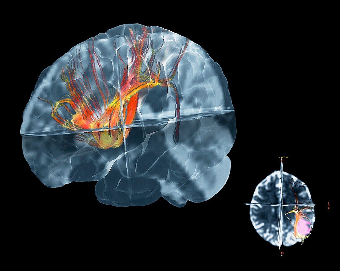
| 3-D Coloured Tractography Magnetic Resonance Imaging (MRI) brain scan of a patient aged 36 with an oligodendroglioma brain tumour (cancer,at centre) in the parietal region of the left hemisphere of the cerebrum. This cancer does not spread but damages the brain as it grows. Here,this tractography scan reveals how nerve fibres (red,yellow) have been affected by the tumour,in particular how one nerve fibre tract arcs around the tumour then turns downwards. The scan at lower right shows the tumour (pink) and nerve fibres deviating around it | |
| Licence : | Droits gérés |
| Crédit: | Science Photo Library / Zephyr |
| Taille de l’image : | 4706 px × 3744 px |
| Model Release : | Non requis |
| Property Release : | Non requis |
| Restrictions : | - |
Prix pour cette image À partir de 45 €
Produit vendu
(Calendrier, Carte postale, Carte de vœux, Impression sur textile, Packaging etc)
À partir de 45 €
Usage commercial
(Affichage, Annonce presse, Annonce TV, Carte, Digital - hors rés. sociaux, Digital - rés. sociaux etc)
À partir de 45 €
Éditorial
(Digital, Journal, Livre, Livre pratique, Magazine, Télévision etc)
À partir de 60 €
Usage non-commercial
(Digital - hors rés. sociaux, Digital - rés. sociaux etc)
À partir de 120 €
Mots clés
- 3 D,
- 3 dimensions,
- 3-D,
- 3D,
- cancer,
- cérébral,
- cerebrum,
- cerveau,
- désordre,
- en 3D,
- fibres nerveuses,
- glioma,
- gliome,
- hémisphère gauche,
- imagerie par résonance magnétique,
- lobe pariétal,
- maladie,
- médical,
- médicale,
- neuro-imagerie,
- neuroimagerie,
- oligodendrogliome,
- soins de santé,
- tri-dimensionnel,
- tridimensionnel,
- trois dimensions,
- trouble,
- tumeur