Deer antler,SEM
Numéro d’image : 11559295
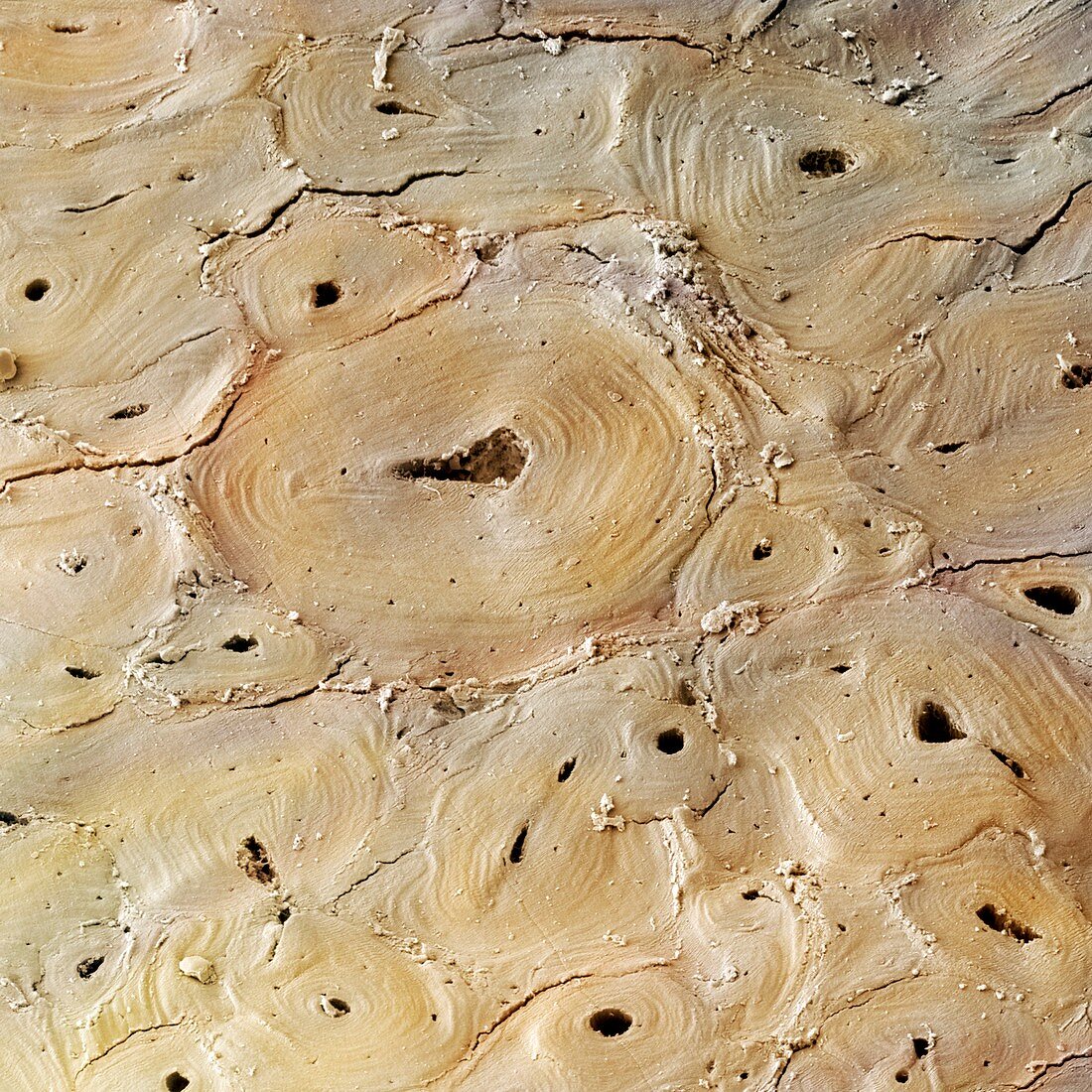
| Deer antler. Coloured scanning electron micrograph (SEM) of a transverse section through cortical (compact) bone from the antler of a deer. The larger spaces seen here are Haversian canals,which contain blood and lymph vessels and nerves. The smaller spaces,known as lacunae,house the osteocytes,the bone-forming cells. Antlers consist entirely of bone and are shed every year. Magnification: x200 when printed at 10 centimetres across | |
| Licence : | Droits gérés |
| Crédit: | Science Photo Library / Power And Syred |
| Taille de l’image : | 4800 px × 4800 px |
| Model Release : | Non requis |
| Property Release : | Non requis |
| Restrictions : | - |
Prix pour cette image À partir de 45 €
Produit vendu
(Calendrier, Carte postale, Carte de vœux, Impression sur textile, Packaging etc)
À partir de 45 €
Usage commercial
(Affichage, Annonce presse, Annonce TV, Carte, Digital - hors rés. sociaux, Digital - rés. sociaux etc)
À partir de 45 €
Éditorial
(Digital, Journal, Livre, Livre pratique, Magazine, Télévision etc)
À partir de 60 €
Usage non-commercial
(Digital - hors rés. sociaux, Digital - rés. sociaux etc)
À partir de 120 €
Mots clés
- andouiller,
- animal,
- biologie,
- biologique,
- bois,
- canal Haversian,
- canaux,
- catégorie,
- cerf,
- collagène,
- coloré,
- colorié,
- colorisé,
- coupe,
- divisé,
- en bonne santé,
- faune,
- Lacunae,
- lacune,
- lamella,
- lamelle,
- M.E.B.,
- matrice,
- MEB,
- microscope électronique à balayage,
- nature,
- normal,
- os compact,
- os cortical,
- ostéocyte,
- partie,
- sain,
- section,
- structure,
- transversal,
- transversale,
- zoologie,
- zoologique