Leaf section,SEM
Numéro d’image : 11551351
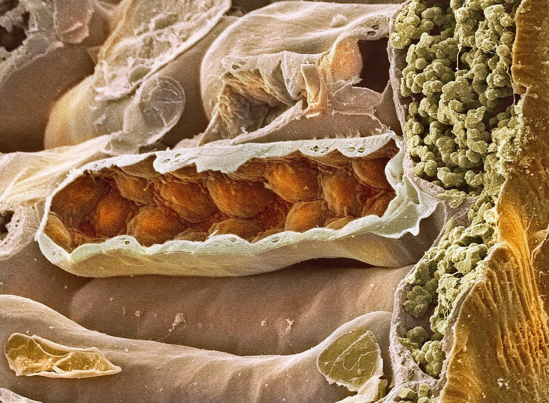
| Leaf section. Coloured scanning electron micrograph (SEM) of a section through a fractured leaf. At right is a single layer of cells that forms the epidermis of the leaf. The top layer (seen here at left) of the leaf interior is made up of palisade mesophyll tissue. One of the tightly packed palisade cells (light brown) has been fractured open to reveal its chloroplasts (dark brown),the sites of photosynthesis | |
| Licence : | Droits gérés |
| Crédit: | Science Photo Library / Furness, Dr. David |
| Taille de l’image : | 4911 px × 3602 px |
| Model Release : | Non requis |
| Property Release : | Non requis |
| Restrictions : | - |
Prix pour cette image À partir de 45 €
Produit vendu
(Calendrier, Carte postale, Carte de vœux, Impression sur textile, Packaging etc)
À partir de 45 €
Usage commercial
(Affichage, Annonce presse, Annonce TV, Carte, Digital - hors rés. sociaux, Digital - rés. sociaux etc)
À partir de 45 €
Éditorial
(Digital, Journal, Livre, Livre pratique, Magazine, Télévision etc)
À partir de 60 €
Usage non-commercial
(Digital - hors rés. sociaux, Digital - rés. sociaux etc)
À partir de 120 €
Mots clés
- anatomie végétale,
- biologie,
- biologique,
- botanique,
- cellule,
- cellules,
- chloroplaste,
- chloroplastes,
- coloré,
- colorié,
- colorisé,
- coupe transversale,
- divisé,
- épiderme,
- flore,
- M.E.B.,
- MEB,
- microscope électronique à balayage,
- nature,
- organite,
- palisade mesophyll,
- photosynthèse,
- plante,
- structure,
- tissus,
- vert