Graafian follicle,light micrograph
Numéro d’image : 11549633
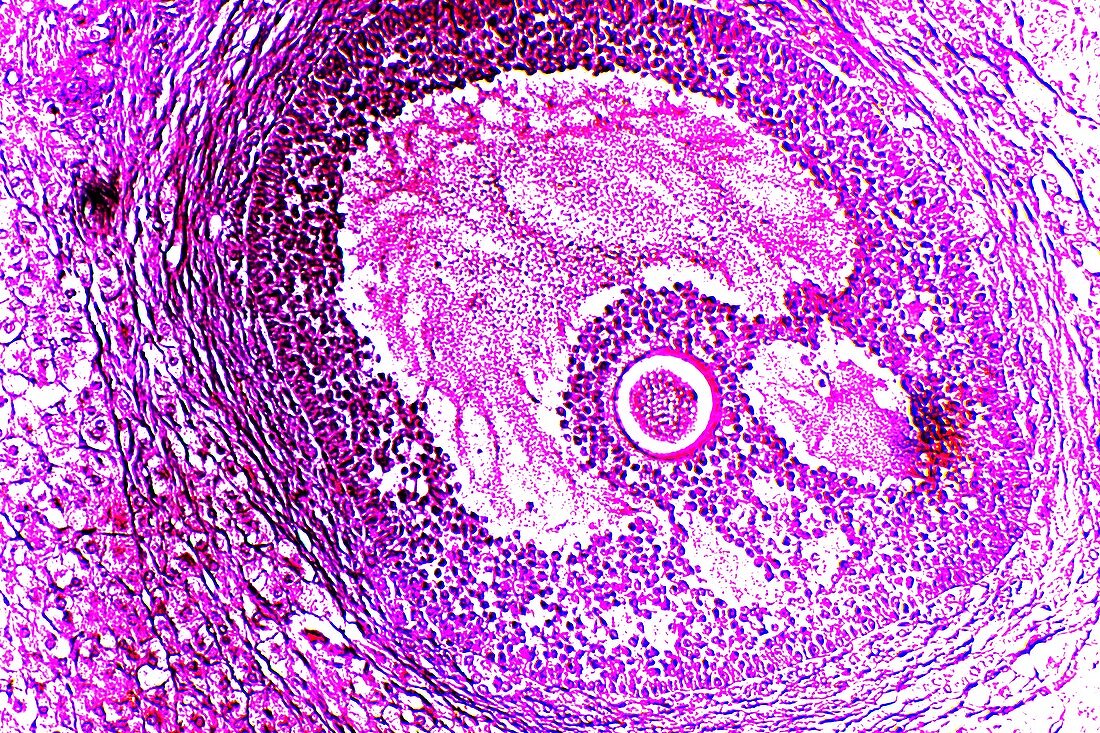
| Graafian follicle. Light micrograph of a section through a mature Graafian follicle from a mammalian ovary. This contains the secondary oocyte (round object at centre),which becomes an ovum (egg cell) when it is released into the oviduct (fallopian tube). The secondary oocyte is surrounded by cells (corona radiata) within the cavity of the follicle,which is filled with follicular fluid. The follicle wall (outer area) consists of an inner layer (membrana granulosa) and an outer fibrous layer. Magnification: x103 when printed at 10 centimetres across | |
| Licence : | Droits gérés |
| Crédit: | Science Photo Library / Wheeler, Dr. Keith |
| Taille de l’image : | 5616 px × 3744 px |
| Model Release : | Non requis |
| Property Release : | Non requis |
| Restrictions : | - |
Prix pour cette image À partir de 45 €
Produit vendu
(Calendrier, Carte postale, Carte de vœux, Impression sur textile, Packaging etc)
À partir de 45 €
Usage commercial
(Affichage, Annonce presse, Annonce TV, Carte, Digital - hors rés. sociaux, Digital - rés. sociaux etc)
À partir de 45 €
Éditorial
(Digital, Journal, Livre, Livre pratique, Magazine, Télévision etc)
À partir de 60 €
Usage non-commercial
(Digital - hors rés. sociaux, Digital - rés. sociaux etc)
À partir de 120 €
Mots clés
- anatomie,
- anatomique,
- biologie,
- biologique,
- catégorie,
- corona radiata,
- coupe,
- divisé,
- en bonne santé,
- follicule,
- follicule de Graaf,
- follicules,
- histologie,
- histologique,
- mammifère,
- mauve,
- micrographie optique,
- microscope optique,
- microscopie optique,
- normal,
- oocute,
- ovaire,
- ovarien,
- ovocyte,
- ovule humain,
- partie,
- personne,
- pourpre,
- reproduction,
- sain,
- section,
- système reproducteur féminin,
- tissus,
- violet