Single-cell desmid,Micrasterias
Numéro d’image : 11526479
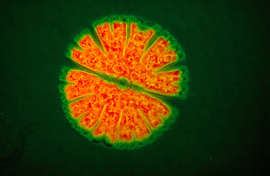
| Ultraviolet fluorescence light micrograph of a single- cell desmid Micrasterias sp. The cell is divided into two halves,separated by a narrow waist. Division of the desmid occurs by splitting in two at the waist,with each half generating a perfect replica of itself to restore the original shape. When exposed to ultraviolet light,chlorophyll,the light sensitive pigment contained within green algae,fluoresces intensely. The red coloured areas in this micrograph correspond to chlorophyll in the cell | |
| Licence : | Droits gérés |
| Crédit: | Science Photo Library / Hinsch, Jan |
| Taille de l’image : | 3798 px × 2480 px |
| Model Release : | Non requis |
| Property Release : | Non requis |
| Restrictions : | - |
Prix pour cette image À partir de 45 €
Produit vendu
(Calendrier, Carte postale, Carte de vœux, Impression sur textile, Packaging etc)
À partir de 45 €
Usage commercial
(Affichage, Annonce presse, Annonce TV, Carte, Digital - hors rés. sociaux, Digital - rés. sociaux etc)
À partir de 45 €
Éditorial
(Digital, Journal, Livre, Livre pratique, Magazine, Télévision etc)
À partir de 60 €
Usage non-commercial
(Digital - hors rés. sociaux, Digital - rés. sociaux etc)
À partir de 120 €