Fungus Penicillium sp. condiophores
Numéro d’image : 11525303
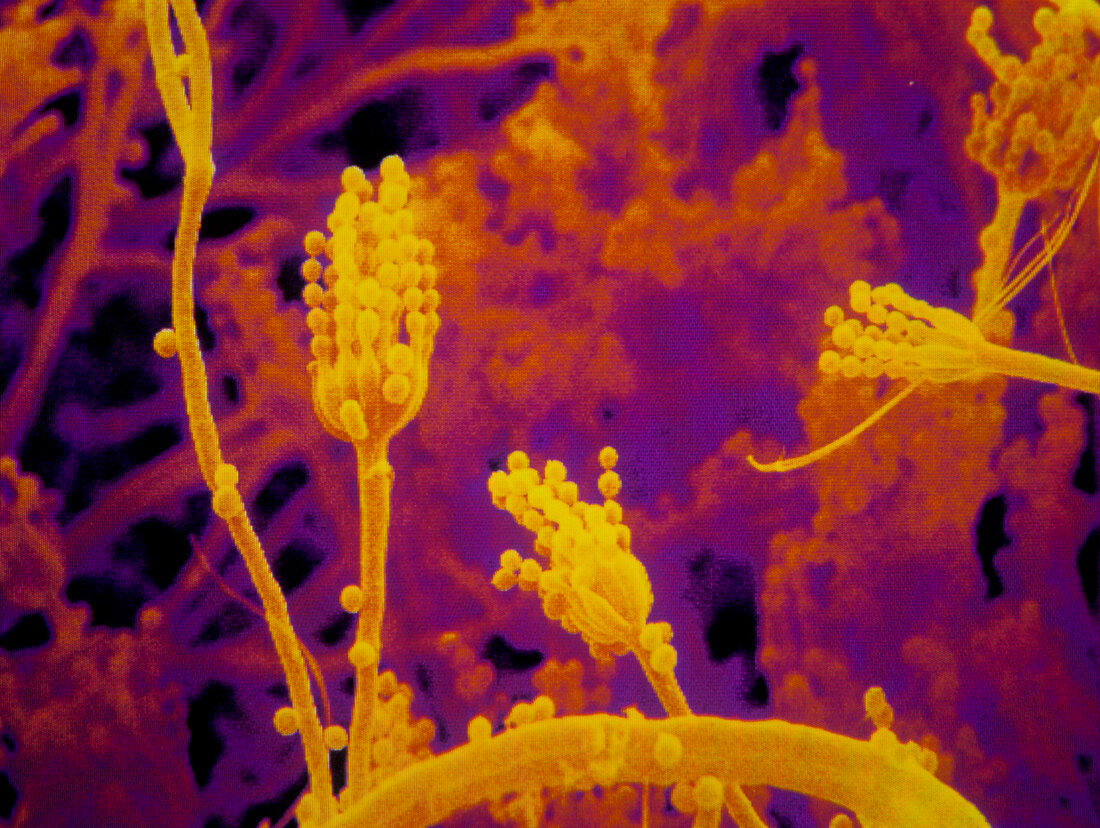
| Fungus. Coloured scanning electron micrograph of a Penicillium sp. fungus. In this image,the condiophores are seen. They are the stalk-like objects,to which are attached numerous round condia. The conidia are the fruiting bodies of the fungus. Magnification: x900 at 6x4.5cm size | |
| Licence : | Droits gérés |
| Crédit: | Science Photo Library / Power And Syred |
| Taille de l’image : | 3552 px × 2674 px |
| Model Release : | Non requis |
| Property Release : | Non requis |
| Restrictions : | - |
Prix pour cette image À partir de 45 €
Produit vendu
(Calendrier, Carte postale, Carte de vœux, Impression sur textile, Packaging etc)
À partir de 45 €
Usage commercial
(Affichage, Annonce presse, Annonce TV, Carte, Digital - hors rés. sociaux, Digital - rés. sociaux etc)
À partir de 45 €
Éditorial
(Digital, Journal, Livre, Livre pratique, Magazine, Télévision etc)
À partir de 60 €
Usage non-commercial
(Digital - hors rés. sociaux, Digital - rés. sociaux etc)
À partir de 120 €