False-colour SEM of the fungus Fusarium oxysporum
Numéro d’image : 11525271
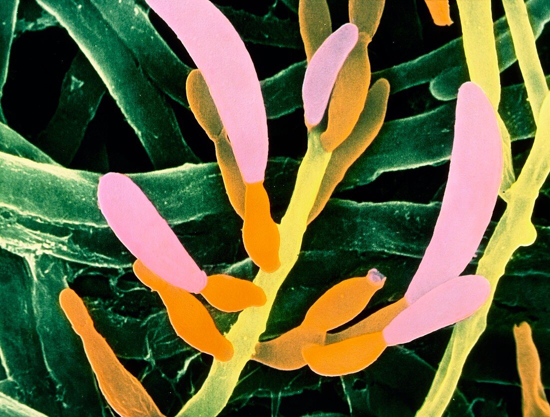
| False-colour scanning electron micrograph of the pathogenic fungus Fusarium oxysporum,which causes wilt disease in tomato and carnation plants. The micrograph shows the hyphae,or vegetative structure of the fungus in the background coloured green. The pink and organge structures,branching off from main hyphal filaments,are conidiophores or specialised spore producing bodies. The conidia,or spores,are produced from the tips of these structures and when ripe are released into the soil. One such structure that has released its conidia is visible at top right showing a jagged crown-like opening. Magnification: x2000 at 6x4.4cm size | |
| Licence : | Droits gérés |
| Crédit: | Science Photo Library / Burgess, Dr. Jeremy |
| Taille de l’image : | 4829 px × 3661 px |
| Model Release : | Non requis |
| Property Release : | Non requis |
| Restrictions : | - |
Prix pour cette image À partir de 45 €
Produit vendu
(Calendrier, Carte postale, Carte de vœux, Impression sur textile, Packaging etc)
À partir de 45 €
Usage commercial
(Affichage, Annonce presse, Annonce TV, Carte, Digital - hors rés. sociaux, Digital - rés. sociaux etc)
À partir de 45 €
Éditorial
(Digital, Journal, Livre, Livre pratique, Magazine, Télévision etc)
À partir de 60 €
Usage non-commercial
(Digital - hors rés. sociaux, Digital - rés. sociaux etc)
À partir de 120 €