LM of a monolayer of onion cells
Numéro d’image : 11523594
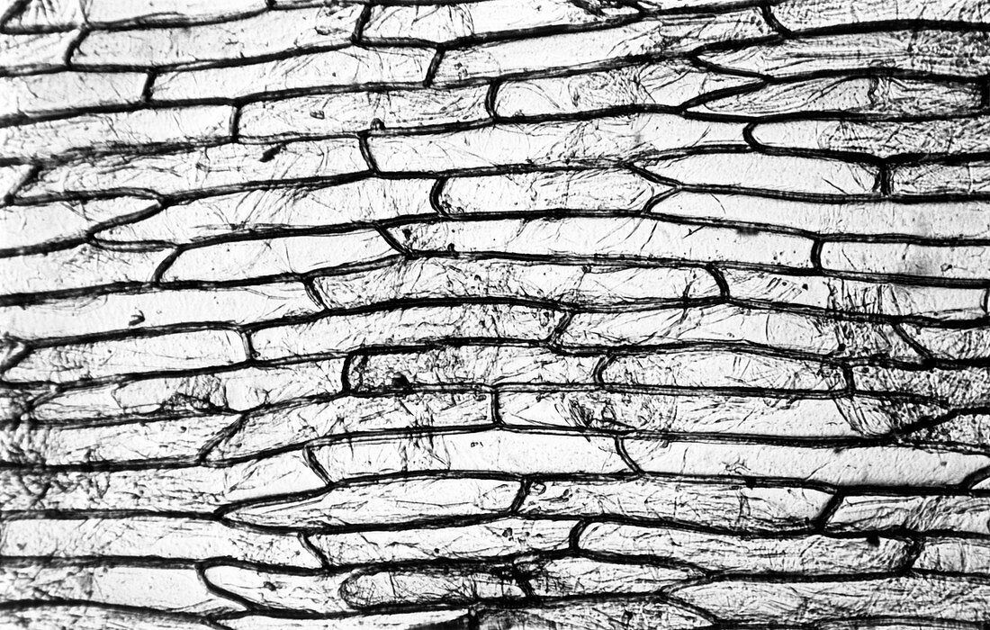
| Light micrograph of a monolayer of cells from an onion Allium cepa,showing the elongated shape of the individual cells. The cell walls,which appear as dark outlines,are wedged close to each other in neat order. This microscope slide was made as an example of a normal preparation technique for use under a light microscope. It compares with B060/050 through B060/053,which were made to show poor techniques,such as the inclusion of air bubbles on the slide or folded layers of tissue that obscure details of the cells. Magnification: x100 at 8x10 inch size | |
| Licence : | Droits gérés |
| Crédit: | Science Photo Library / Stammers, Sinclair |
| Taille de l’image : | 3898 px × 2480 px |
| Model Release : | Non requis |
| Property Release : | Non requis |
| Restrictions : | - |
Prix pour cette image À partir de 45 €
Produit vendu
(Calendrier, Carte postale, Carte de vœux, Impression sur textile, Packaging etc)
À partir de 45 €
Usage commercial
(Affichage, Annonce presse, Annonce TV, Carte, Digital - hors rés. sociaux, Digital - rés. sociaux etc)
À partir de 45 €
Éditorial
(Digital, Journal, Livre, Livre pratique, Magazine, Télévision etc)
À partir de 60 €
Usage non-commercial
(Digital - hors rés. sociaux, Digital - rés. sociaux etc)
À partir de 120 €