Light micrograph of a fresh tulip petal
Numéro d’image : 11523577
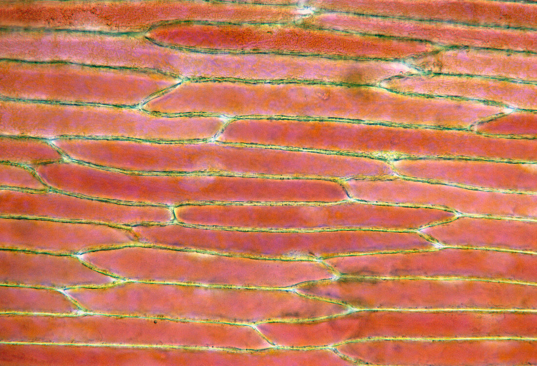
| Light micrograph of a tulip petal. This photograph shows the cells of a fresh petal bounded by cell walls (yellow),with cytoplasm (orange,natural colour) filling each of the cells. As the petal begins to wilt,water is lost and the cytoplasm retracts into the centre of each cell. Photo number B060/023 shows a wilted petal from the same flower. Magnification: x150 at 35mm size | |
| Licence : | Droits gérés |
| Crédit: | Science Photo Library / Nuridsany, Claude / Perennou, Maria |
| Taille de l’image : | 5114 px × 3485 px |
| Model Release : | Non requis |
| Property Release : | Non requis |
| Restrictions : |
|
Prix pour cette image À partir de 45 €
Produit vendu
(Calendrier, Carte postale, Carte de vœux, Impression sur textile, Packaging etc)
À partir de 45 €
Usage commercial
(Affichage, Annonce presse, Annonce TV, Carte, Digital - hors rés. sociaux, Digital - rés. sociaux etc)
À partir de 45 €
Éditorial
(Digital, Journal, Livre, Livre pratique, Magazine, Télévision etc)
À partir de 60 €
Usage non-commercial
(Digital - hors rés. sociaux, Digital - rés. sociaux etc)
À partir de 120 €