Surface of protoplast from tobacco leaf
Numéro d’image : 11523574
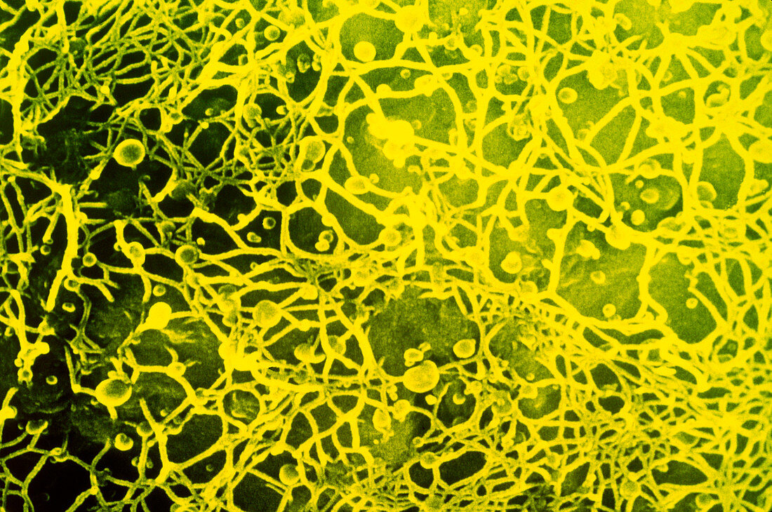
| False-colour scanning electron micrograph of details of the surface of a tobacco leaf protoplast,Nicotiana tabacum (cv White Burley) showing the regrowth of the cell wall ten hours after culturing. The fibres visible correspond to cellulose microfibrils,beneath is the plasma membrane and this highlights the underlying chloroplasts. The spherical bodies visible among the fibres are either membrane blebs or artefacts. Magnification: x3430 at 35mm size | |
| Licence : | Droits gérés |
| Crédit: | Science Photo Library / Burgess, Dr. Jeremy |
| Taille de l’image : | 5083 px × 3373 px |
| Model Release : | Non requis |
| Property Release : | Non requis |
| Restrictions : | - |
Prix pour cette image À partir de 45 €
Produit vendu
(Calendrier, Carte postale, Carte de vœux, Impression sur textile, Packaging etc)
À partir de 45 €
Usage commercial
(Affichage, Annonce presse, Annonce TV, Carte, Digital - hors rés. sociaux, Digital - rés. sociaux etc)
À partir de 45 €
Éditorial
(Digital, Journal, Livre, Livre pratique, Magazine, Télévision etc)
À partir de 60 €
Usage non-commercial
(Digital - hors rés. sociaux, Digital - rés. sociaux etc)
À partir de 120 €