Model of the protein actin
Numéro d’image : 11521398
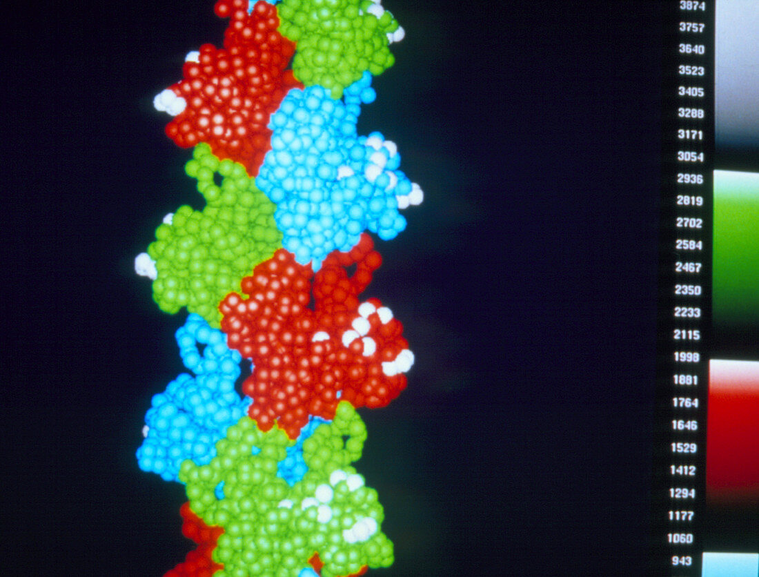
| Computer generated space-filling model showing ten actin monomers from an F-actin helix,a polymer of the protein actin. For clarity each actin monomer (the repeated polymer unit) is shown in a different colour. Each amino-acid residue is represented by a sphere of radius 2.7 angstroms. In muscle cells,actin forms part of the thin filament,which cyclically interacts with the thick myosin filament to produce a mutual sliding that is the basis of muscle contraction. The white spheres are amino-acid residues that cross-link to the myosin in the actomyosin complex. The structure of the F-actin filament was determined using a technique called X-ray fibre diffraction | |
| Licence : | Droits gérés |
| Crédit: | Science Photo Library / Holmes, Dr. Kenneth |
| Taille de l’image : | 3030 px × 2303 px |
| Model Release : | Non requis |
| Property Release : | Non requis |
| Restrictions : | - |
Prix pour cette image À partir de 45 €
Produit vendu
(Calendrier, Carte postale, Carte de vœux, Impression sur textile, Packaging etc)
À partir de 45 €
Usage commercial
(Affichage, Annonce presse, Annonce TV, Carte, Digital - hors rés. sociaux, Digital - rés. sociaux etc)
À partir de 45 €
Éditorial
(Digital, Journal, Livre, Livre pratique, Magazine, Télévision etc)
À partir de 60 €
Usage non-commercial
(Digital - hors rés. sociaux, Digital - rés. sociaux etc)
À partir de 120 €