Human serum albumin
Numéro d’image : 11521393
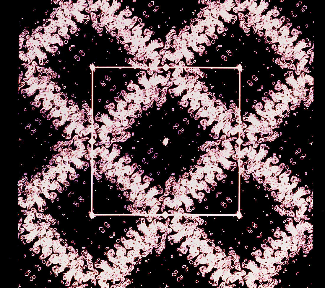
| Cross section of a computerised model of a crystal of Human Serum Albumin (each side of a square represents one molecule). Images are made by X- raying a crystal of the protein and recording a projection of its electron density. By stacking several such cross-sections together,a low resolution 3-dimensional model of the protein may be built. Knowledge of the molecular structure,including how and where substrates such as minerals and therapeutic drugs bind on,should open the way for pharmaceutical companies to design new drugs or alter existing ones to allow them to be carried more efficiently by the protein molecule through the body | |
| Licence : | Droits gérés |
| Crédit: | Science Photo Library / NASA |
| Taille de l’image : | 4451 px × 3957 px |
| Model Release : | Non requis |
| Property Release : | Non requis |
| Restrictions : | - |
Prix pour cette image À partir de 45 €
Produit vendu
(Calendrier, Carte postale, Carte de vœux, Impression sur textile, Packaging etc)
À partir de 45 €
Usage commercial
(Affichage, Annonce presse, Annonce TV, Carte, Digital - hors rés. sociaux, Digital - rés. sociaux etc)
À partir de 45 €
Éditorial
(Digital, Journal, Livre, Livre pratique, Magazine, Télévision etc)
À partir de 60 €
Usage non-commercial
(Digital - hors rés. sociaux, Digital - rés. sociaux etc)
À partir de 120 €
Mots clés
- albumine,
- biochimie,
- biochimique,
- chimie,
- chimique,
- composé,
- composés,
- conception de médicament,
- cristallographie au rayon X,
- diffractométrie de rayon X,
- DRX,
- éléments,
- graphiques informatiques,
- infographies,
- molécule,
- molécules,
- personne,
- produit sanguin,
- protéine,
- protéines,
- radiocristallographie,
- radiographie,
- rayons X,
- sérum albumine,
- sérum-albumine,
- XRD