Images
Vidéos
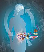
13839367 - Gut brain connection, illustration
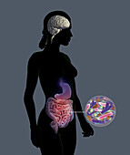
13839363 - Gut brain connection, illustration

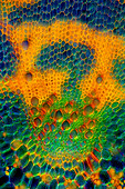
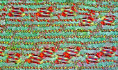
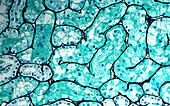
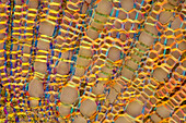
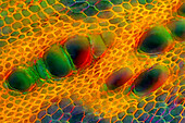
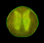
13838447 - Reed stalk, light micrograph
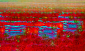
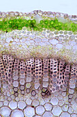
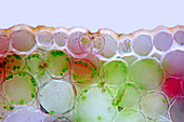
13838403 - Nettle stalk, light micrograph
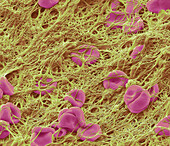
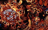
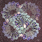
13756293 - Blood clot, SEM

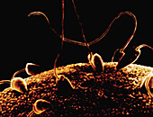
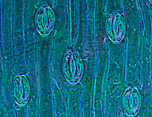
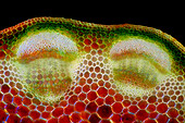
13755913 - Oat stoma, light micrograph
13755912 - Oat stomata, light micrograph
13755907 - Oat stomata, light micrograph
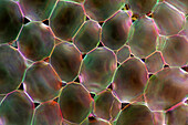
13755899 - Narrowleaf lupin stalk, light micrograph
13755898 - Narrowleaf lupin stalk, light micrograph
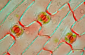
13755868 - Lily of the valley stomata, light micrograph
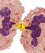
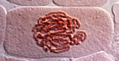
13755100 - Mitosis, light micrograph
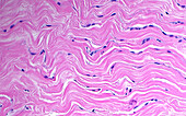
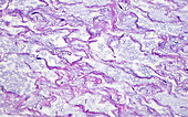
13732520 - Collagen fibres in breast tissue, light micrograph
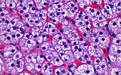
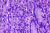
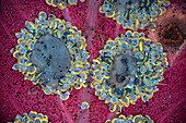
13732517 - Adrenal gland cortex cells, light micrograph
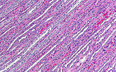
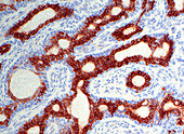
13732477 - Kidney medulla, light micrograph
13732444 - Stomach adenocarcinoma cells, light micrograph
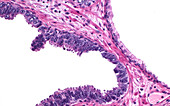
13732439 - Prostate intraepithelial neoplasia, light micrograph
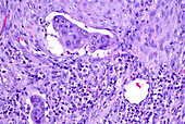
13732413 - Cancer cells blood vessel invasion, light micrograph
13732368 - Kidney proximal convoluted tubules, light micrograph
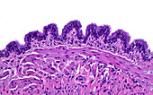
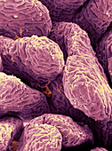
13732352 - Respiratory tract lining cells and cilia, light micrograph
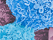
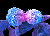
13672040 - Lung cancer cells dividing, SEM
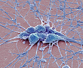
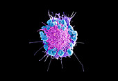
13671965 - Neurones, SEM
13633941 - Field scabious stalk, light micrograph
13632808 - Reflex arc, illustration
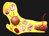
13632786 - Fertilisation, illustration
13632784 - Blood cell production, illustration

13613778 - Haemocytometer
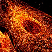
13243195 - Cancer cell showing tubulin, light micrograph
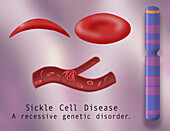
14078387 - Normal blood cell and sickle cell disease, illustration
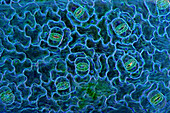
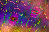
13954538 - Stomata in tulip leaf epidermis, light micrograph
13954442 - Stomata in spath leaf epidermis, light micrograph
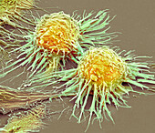
13952286 - Prostate cancer cells dividing, SEM
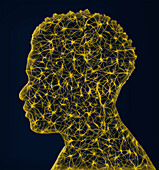
13950834 - Neuron head, conceptual illustration
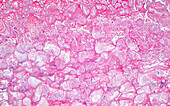
13950511 - Saponification fat necrosis, light micrograph
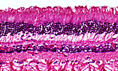
13950466 - Human retina cells, light micrograph
13950387 - Newt larvae ciliated cells, SEM
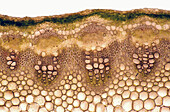
13838435 - Pennycress stalk, light micrograph

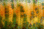
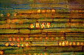
13838420 - Senecio sp. vascular bundle, light micrograph
13838409 - Nettle stalk, light micrograph
13765243 - Tooth bacteria, SEM
13765236 - Schizosaccharomyces pombe yeast, SEM
13755870 - Lily of the valley stomata, light micrograph
13755865 - Lily of the valley stomata, light micrograph
13755853 - Marsh-marigold leaf, light micrograph
13755839 - Mitosis, light micrograph
13755592 - Mitosis, light micrograph
13733329 - Cervical cancer cells dividing, SEM
13732928 - Aspergillus fungus, SEM
13732505 - Umbilical cord substance, light micrograph

13732424 - Prostate corpus amylaceous, light micrograph

13732404 - Bladder cancer invasion, light micrograph
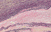
13732374 - Aorta aneurysm elastic stain, light micrograph
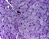
13686213 - Skeletal muscle fibres, light micrograph
13685621 - Neuromuscular junction, TEM
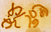
13674116 - Anabaena sp. cyanobacteria, light micrograph
13672036 - Cervical cancer cell, SEM
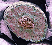
13634170 - Testis, SEM
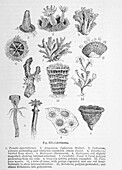
13634097 - Cells under microscope, 19th century illustration

13632807 - Fertilisation, illustration
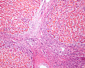
13601241 - Cirrhosis, light micrograph
14084178 - Vaginitis, LM
14084172 - Nucleated RBCs in placental, LM
14084164 - Vaginitis, LM
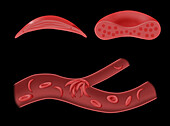
14078377 - Normal blood cell and sickle cell disease, illustration
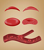
14078375 - Normal blood cell and sickle cell disease, illustration
13954455 - Dividing cancer cell, TEM
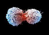
13952288 - Prostate cancer cells dividing, SEM
13950531 - Fabry’s disease lamellar bodies, TEM
13950530 - Woven bone, light micrograph
13950455 - Glomerulus crescent, light micrograph
13950425 - Pleomorphic adenoma matrix, light micrograph
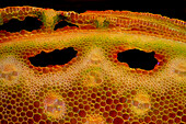
13838444 - Reed stalk, light micrograph
13838423 - Knautia sp. stalk, light micrograph
13838418 - Fleabane stalk, light micrograph
13838411 - Nettle stalk, light micrograph

13838406 - Nettle stalk, light micrograph
13838404 - Nettle stalk, light micrograph

13756607 - Cervical cancer cells dividing, SEM
13755873 - Lily of the valley leaf epidermis, light micrograph
13755862 - Red clover stalk, light micrograph
13755802 - Ciliary doublet microtubule, illustration
13754707 - Mitosis, light micrograph
13732529 - Basal cell adenoma immunostain, light micrograph
13686771 - Fern sorus and spore cases, fluorescence light micrograph
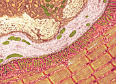
13686445 - Small intestine, SEM
13686367 - Spinal cord, light micrograph

13686362 - Respiratory epithelium, TEM
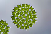
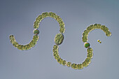
13674090 - Green alga, light micrograph
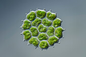
13674038 - Green alga, light micrograph
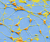
13633227 - Pluripotent derived neurones, SEM
13601227 - Fatty liver disease, light micrograph
14084187 - Breast cancer, LM
14084171 - Nucleated RBCs in placental, LM
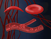
14078386 - Normal blood cell and sickle cell disease, illustration
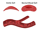
14078378 - Normal blood cell and sickle cell disease, illustration
14077660 - Dividing HeLa cells, SEM
13954448 - Stomata in spath leaf epidermis, light micrograph
13954393 - Stomata in croton leaf, light micrograph

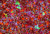
13951659 - Behaviour of red blood cell in hypotonic medium, illustration
page suivante
Cellules Photos ❘ Science Photo Library