Human aorta, light micrograph
Numéro d’image : 13613431
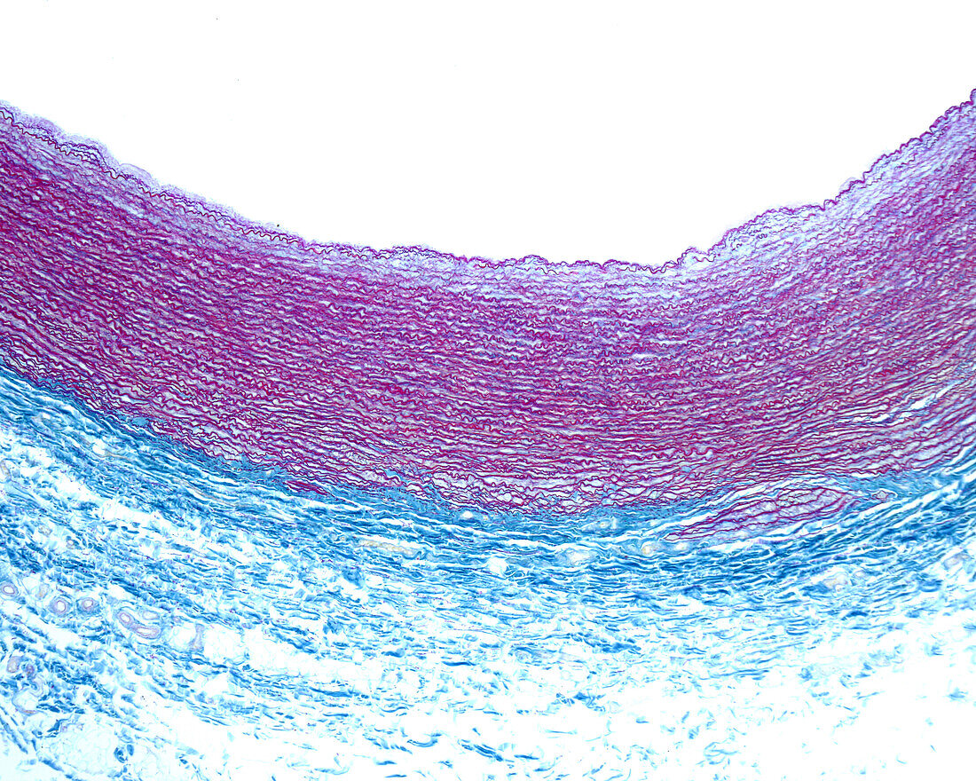
| Light micrograph of a cross-sectioned wall of a human aorta stained with the Cajal-Gallego method for the demonstration of elastic elements. The method allows a clear distinction between the tunica media with abundant elastic lamellae (stained magenta by fuchsin) and the tunica adventitia, which is rich in collagen fibres (stained blue by indigo carmine). | |
| Licence : | Droits gérés |
| Crédit: | Science Photo Library / JOSE CALVO |
| Taille de l’image : | 3840 px × 3072 px |
| Model Release : | Non requis |
| Property Release : | Non requis |
| Restrictions : | - |
Prix pour cette image À partir de 45 €
Produit vendu
(Calendrier, Carte postale, Carte de vœux, Impression sur textile, Packaging etc)
À partir de 45 €
Usage commercial
(Affichage, Annonce presse, Annonce TV, Carte, Digital - hors rés. sociaux, Digital - rés. sociaux etc)
À partir de 45 €
Éditorial
(Digital, Journal, Livre, Livre pratique, Magazine, Télévision etc)
À partir de 60 €
Usage non-commercial
(Digital - hors rés. sociaux, Digital - rés. sociaux etc)
À partir de 120 €
Mots clés
- adventice,
- adventitatia,
- aorta,
- aorte,
- artère,
- aucun,
- biologie,
- biologique,
- circulatoire,
- élastique,
- en bonne santé,
- histologie,
- histologique,
- humain,
- intima,
- intime,
- médias,
- micrographie optique,
- microscope,
- microscope optique,
- microscopie,
- microscopie optique,
- muscle lisse,
- musculaire,
- normal,
- personne,
- sain,
- tunica,
- vaisseau sanguin,
- vasculaire