Brain haemorrhage, CT scan
Numéro d’image : 13599597
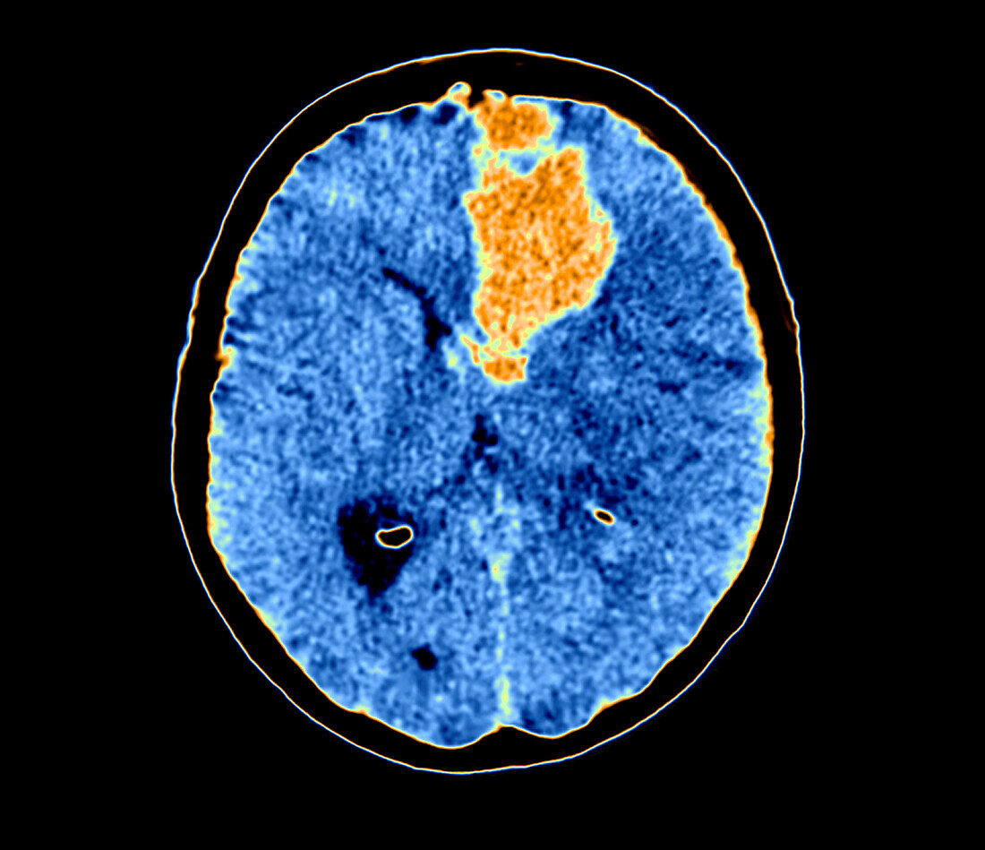
| Coloured axial computed tomography (CT) scan of a 37 year old patient with left hemispherical haematoma (light orange), in the fronto-temporal territory and gray nucleus territory. This type of haematoma results from intra-parenchymal vascular shear proving to be compressive with regard to the ventricular structures and the midline. | |
| Licence : | Droits gérés |
| Crédit: | Science Photo Library / Zephyr |
| Taille de l’image : | 3969 px × 3425 px |
| Model Release : | Non requis |
| Property Release : | Non requis |
| Restrictions : | - |
Prix pour cette image À partir de 45 €
Produit vendu
(Calendrier, Carte postale, Carte de vœux, Impression sur textile, Packaging etc)
À partir de 45 €
Usage commercial
(Affichage, Annonce presse, Annonce TV, Carte, Digital - hors rés. sociaux, Digital - rés. sociaux etc)
À partir de 45 €
Éditorial
(Digital, Journal, Livre, Livre pratique, Magazine, Télévision etc)
À partir de 60 €
Usage non-commercial
(Digital - hors rés. sociaux, Digital - rés. sociaux etc)
À partir de 120 €
Mots clés
- 30,
- accident de la route,
- années 30,
- anormal,
- arrière plan noir,
- arrière-plan noir,
- axial,
- cérébral,
- cerveau,
- coloré,
- colorié,
- colorisé,
- diagnostic,
- diagnostique,
- fond noir,
- haematoma,
- haemorrhagia,
- hématome,
- hémorragie,
- malsain,
- médecine,
- médical,
- médicale,
- neurologie,
- neurologique,
- T.D.M.,
- TDM,
- tomodensitométrie,
- tomographie assistée par ordinateur,
- trauma crânien,
- traumatisme crânien